5B2G image
Deposition Date 2016-01-15
Release Date 2016-10-05
Last Version Date 2024-11-13
Entry Detail
PDB ID:
5B2G
Title:
Crystal structure of human claudin-4 in complex with C-terminal fragment of Clostridium perfringens enterotoxin
Biological Source:
Source Organism:
Host Organism:
Method Details:
Experimental Method:
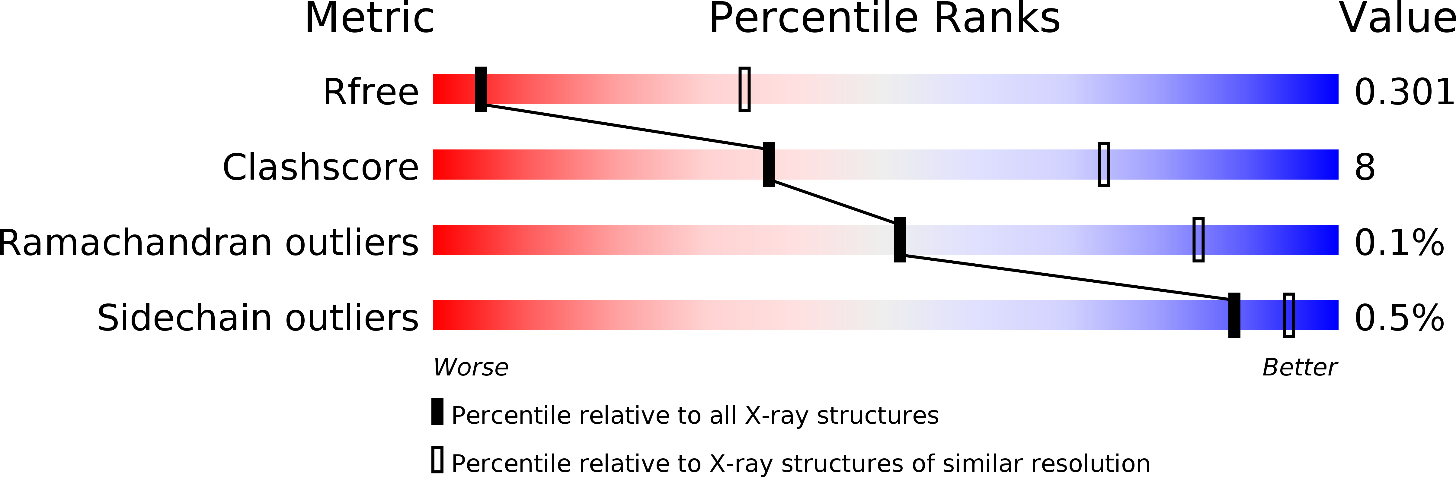
Resolution:
3.50 Å
R-Value Free:
0.30
R-Value Work:
0.28
R-Value Observed:
0.28
Space Group:
P 43
Macromolecular Entities
Protein Blast
Polymer Type:polypeptide(L)
Molecule:Endolysin,Claudin-4
Gene (Uniprot):CLDN4, E
Mutagens:R1012G, C1054T, C1097A, I1137R
Chain IDs:A, C, E, G
Chain Length:364
Number of Molecules:4
Biological Source:Enterobacteria phage T4, Homo sapiens
Structures with similar UniProt ID
Protein Blast
Polymer Type:polypeptide(L)
Molecule:Heat-labile enterotoxin B chain
Gene (Uniprot):cpe
Chain IDs:B, D, F, H
Chain Length:142
Number of Molecules:4
Biological Source:Clostridium perfringens
Modified Residue
Compound ID Chain ID Parent Comp ID Details 2D Image
MSE B MET modified residue
Ligand Molecules
Primary Citation
Structural basis for disruption of claudin assembly in tight junctions by an enterotoxin
Sci Rep 6 33632 33632 (2016)
PMID: 27647526 DOI: 10.1038/srep33632

Abstact

The food-poisoning bacterium Clostridium perfringens produces an enterotoxin (~35 kDa) that specifically targets human claudin-4, among the 26 human claudin proteins, and causes diarrhea by fluid accumulation in the intestinal cavity. The C-terminal domain of the Clostridium perfringens enterotoxin (C-CPE, ~15 kDa) binds tightly to claudin-4, and disrupts the intestinal tight junction barriers. In this study, we determined the 3.5-Å resolution crystal structure of the cell-free synthesized human claudin-4•C-CPE complex, which is significantly different from the structure of the off-target complex of an engineered C-CPE with mouse claudin-19. The claudin-4•C-CPE complex structure demonstrated the mechanism underlying claudin assembly disruption. A comparison of the present C-CPE-bound structure of claudin-4 with the enterotoxin-free claudin-15 structure revealed sophisticated C-CPE-induced conformation changes of the extracellular segments, induced on the foundation of the rigid four-transmembrane-helix bundle structure. These conformation changes provide a mechanistic model for the disruption of the lateral assembly of claudin molecules. Furthermore, the present novel structural mechanism for selecting a specific member of the claudin family can be used as the foundation to develop novel medically important technologies to selectively regulate the tight junctions formed by claudin family members in different organs.

Legend

Protein

Chemical

Disease

Primary Citation of related structures