5B2D image
Entry Detail
PDB ID:
5B2D
Keywords:
Title:
Crystal structure of Mumps virus hemagglutinin-neuraminidase bound to 3-sialyllactose
Biological Source:
Source Organism:
Host Organism:
PDB Version:
Deposition Date:
2016-01-14
Release Date:
2016-09-28
Method Details:
Experimental Method:
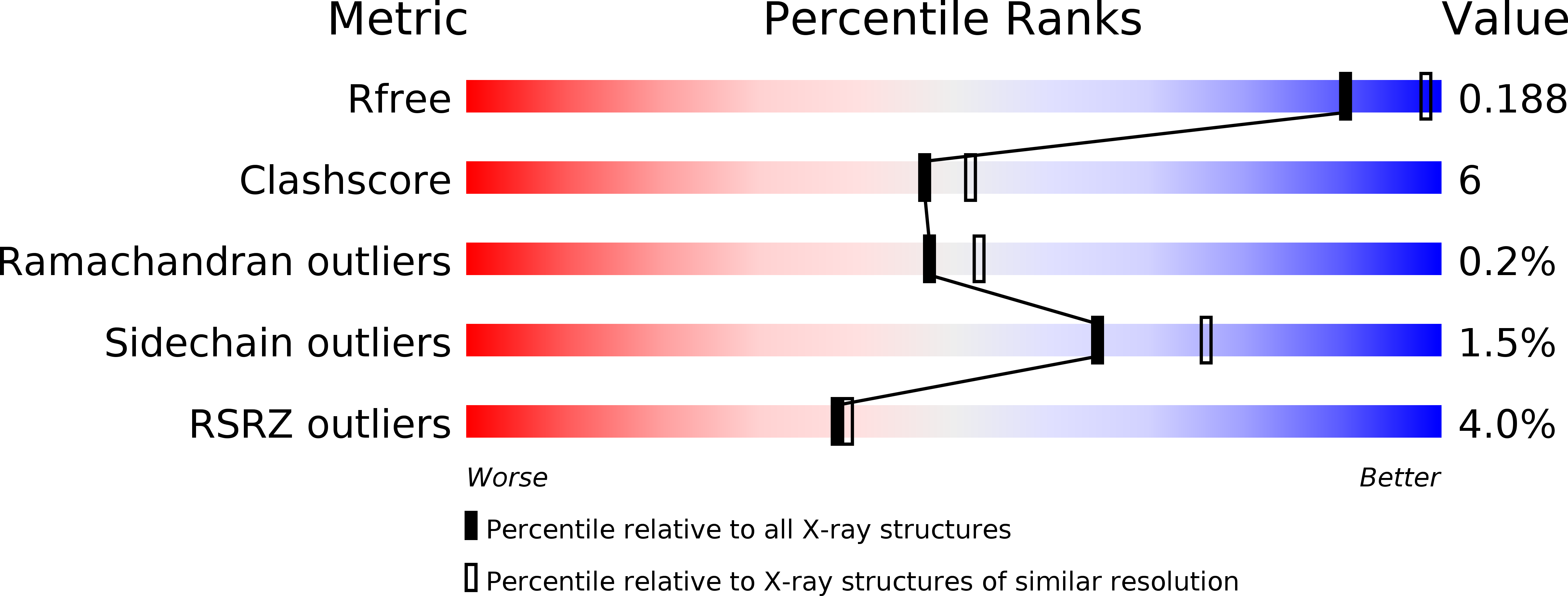
Resolution:
2.18 Å
R-Value Free:
0.19
R-Value Work:
0.17
R-Value Observed:
0.17
Space Group:
P 61
Macromolecular Entities
Polymer Type:polypeptide(L)
Description:HN protein
Chain IDs:A, B
Chain Length:489
Number of Molecules:2
Biological Source:Mumps virus
Peptide-like Molecules
PRD_900025
Primary Citation
Trisaccharide containing alpha 2,3-linked sialic acid is a receptor for mumps virus
Proc.Natl.Acad.Sci.USA 113 11579 11584 (2016)
PMID: 27671656 DOI: 10.1073/pnas.1608383113

Abstact

Mumps virus (MuV) remains an important pathogen worldwide, causing epidemic parotitis, orchitis, meningitis, and encephalitis. Here we show that MuV preferentially uses a trisaccharide containing α2,3-linked sialic acid in unbranched sugar chains as a receptor. Crystal structures of the MuV attachment protein hemagglutinin-neuraminidase (MuV-HN) alone and in complex with the α2,3-sialylated trisaccharide revealed that in addition to the interaction between the MuV-HN active site residues and sialic acid, other residues, including an aromatic residue, stabilize the third sugar of the trisaccharide. The importance of the aromatic residue and the third sugar in the MuV-HN-receptor interaction was confirmed by computational energy calculations, isothermal titration calorimetry studies, and glycan-binding assays. Furthermore, MuV-HN was found to bind more efficiently to unbranched α2,3-sialylated sugar chains compared with branched ones. Importantly, the strategically located aromatic residue is conserved among the HN proteins of sialic acid-using paramyxoviruses, and alanine substitution compromised their ability to support cell-cell fusion. These results suggest that not only the terminal sialic acid but also the adjacent sugar moiety contribute to receptor function for mumps and these paramyxoviruses. The distribution of structurally different sialylated glycans in tissues and organs may explain in part MuV's distinct tropism to glandular tissues and the central nervous system. In the crystal structure, the epitopes for neutralizing antibodies are located around the α-helices of MuV-HN that are not well conserved in amino acid sequences among different genotypes of MuV. This may explain the fact that MuV reinfection sometimes occurs.

Legend

Protein

Chemical

Disease

Primary Citation of related structures