5B24 image
Deposition Date 2015-12-31
Release Date 2016-03-02
Last Version Date 2023-11-08
Entry Detail
PDB ID:
5B24
Title:
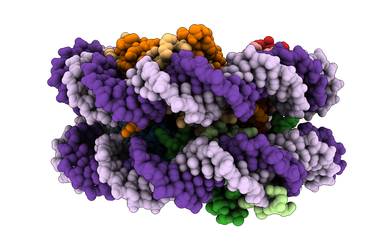
The crystal structure of the nucleosome containing cyclobutane pyrimidine dimer
Biological Source:
Source Organism:
Homo sapiens (Taxon ID: 9606)
Host Organism:
Method Details:
Experimental Method:
Resolution:
3.60 Å
R-Value Free:
0.24
R-Value Work:
0.20
R-Value Observed:
0.20
Space Group:
P 21 21 21
Macromolecular Entities
Structures with similar UniProt ID
Protein Blast
Polymer Type:polypeptide(L)
Molecule:Histone H3.1
Gene (Uniprot):H3C1, H3C2, H3C3, H3C4, H3C6, H3C7, H3C8, H3C10, H3C11, H3C12
Chain IDs:A, E
Chain Length:139
Number of Molecules:2
Biological Source:Homo sapiens
Structures with similar UniProt ID
Protein Blast
Polymer Type:polypeptide(L)
Molecule:Histone H4
Gene (Uniprot):H4C1, H4C2, H4C3, H4C4, H4C5, H4C6, H4C8, H4C9, H4C11, H4C12, H4C13, H4C14, H4C15, H4C16
Chain IDs:B, F
Chain Length:106
Number of Molecules:2
Biological Source:Homo sapiens
Structures with similar UniProt ID
Protein Blast
Polymer Type:polypeptide(L)
Molecule:Histone H2A type 1-B/E
Gene (Uniprot):H2AC4, H2AC8
Chain IDs:C, G
Chain Length:133
Number of Molecules:2
Biological Source:Homo sapiens
Structures with similar UniProt ID
Protein Blast
Polymer Type:polypeptide(L)
Molecule:Histone H2B type 1-J
Gene (Uniprot):H2BC11
Chain IDs:D, H
Chain Length:129
Number of Molecules:2
Biological Source:Homo sapiens
Polymer Type:polydeoxyribonucleotide
Molecule:DNA (145-MER)
Chain IDs:I, J
Chain Length:145
Number of Molecules:2
Biological Source:Homo sapiens
Ligand Molecules
Primary Citation
Crystal structure of the nucleosome containing ultraviolet light-induced cyclobutane pyrimidine dimer
Biochem.Biophys.Res.Commun. 471 117 122 (2016)
PMID: 26837048 DOI: 10.1016/j.bbrc.2016.01.170

Abstact

The cyclobutane pyrimidine dimer (CPD) is induced in genomic DNA by ultraviolet (UV) light. In mammals, this photolesion is primarily induced within nucleosomal DNA, and repaired exclusively by the nucleotide excision repair (NER) pathway. However, the mechanism by which the CPD is accommodated within the nucleosome has remained unknown. We now report the crystal structure of a nucleosome containing CPDs. In the nucleosome, the CPD induces only limited local backbone distortion, and the affected bases are accommodated within the duplex. Interestingly, one of the affected thymine bases is located within 3.0 Å from the undamaged complementary adenine base, suggesting the formation of complementary hydrogen bonds in the nucleosome. We also found that UV-DDB, which binds the CPD at the initial stage of the NER pathway, also efficiently binds to the nucleosomal CPD. These results provide important structural and biochemical information for understanding how the CPD is accommodated and recognized in chromatin.

Legend

Protein

Chemical

Disease

Primary Citation of related structures