Deposition Date
2015-09-27
Release Date
2016-01-13
Last Version Date
2023-11-08
Entry Detail
PDB ID:
5AZ7
Title:
Crystal structure of MBP-Tom20 fusion protein with a 4-residue spacer in the connector helix
Biological Source:
Source Organism(s):
Escherichia coli (strain K12) (Taxon ID: 83333)
Rattus norvegicus (Taxon ID: 10116)
Rattus norvegicus (Taxon ID: 10116)
Expression System(s):
Method Details:
Experimental Method:
Resolution:
1.96 Å
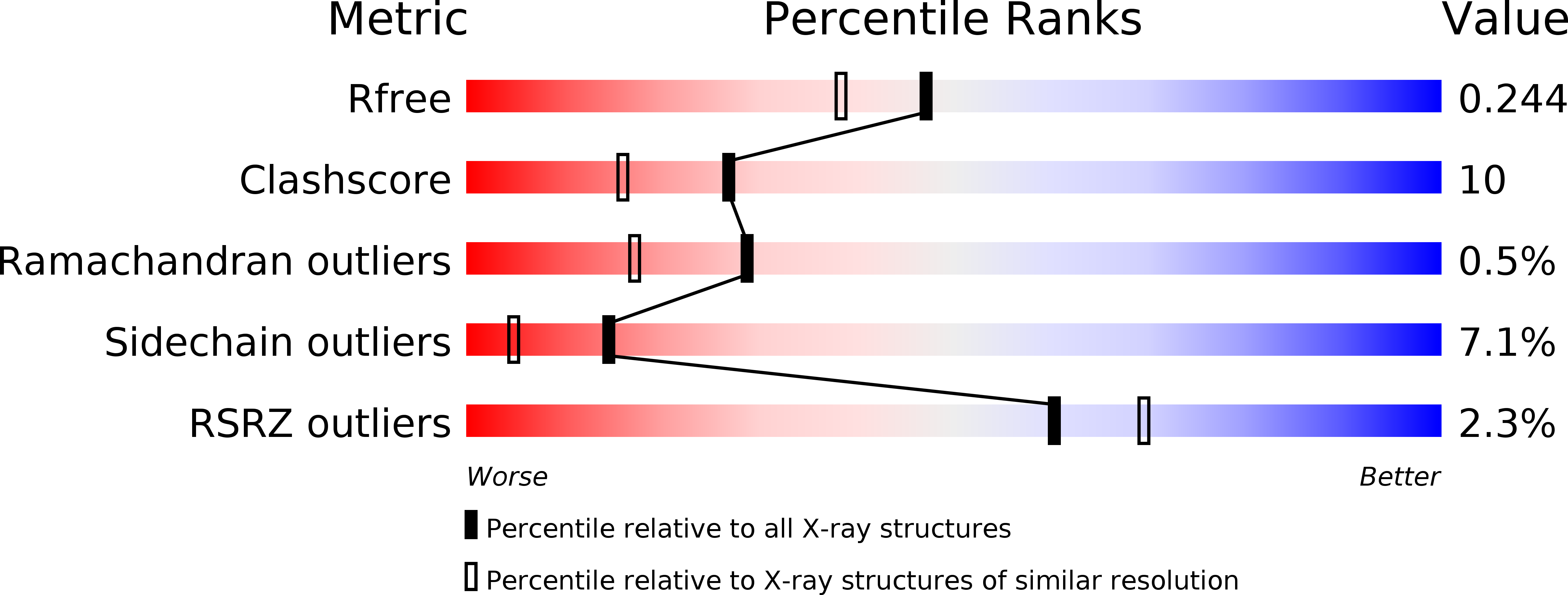
R-Value Free:
0.24
R-Value Work:
0.19
R-Value Observed:
0.19
Space Group:
I 2 2 2