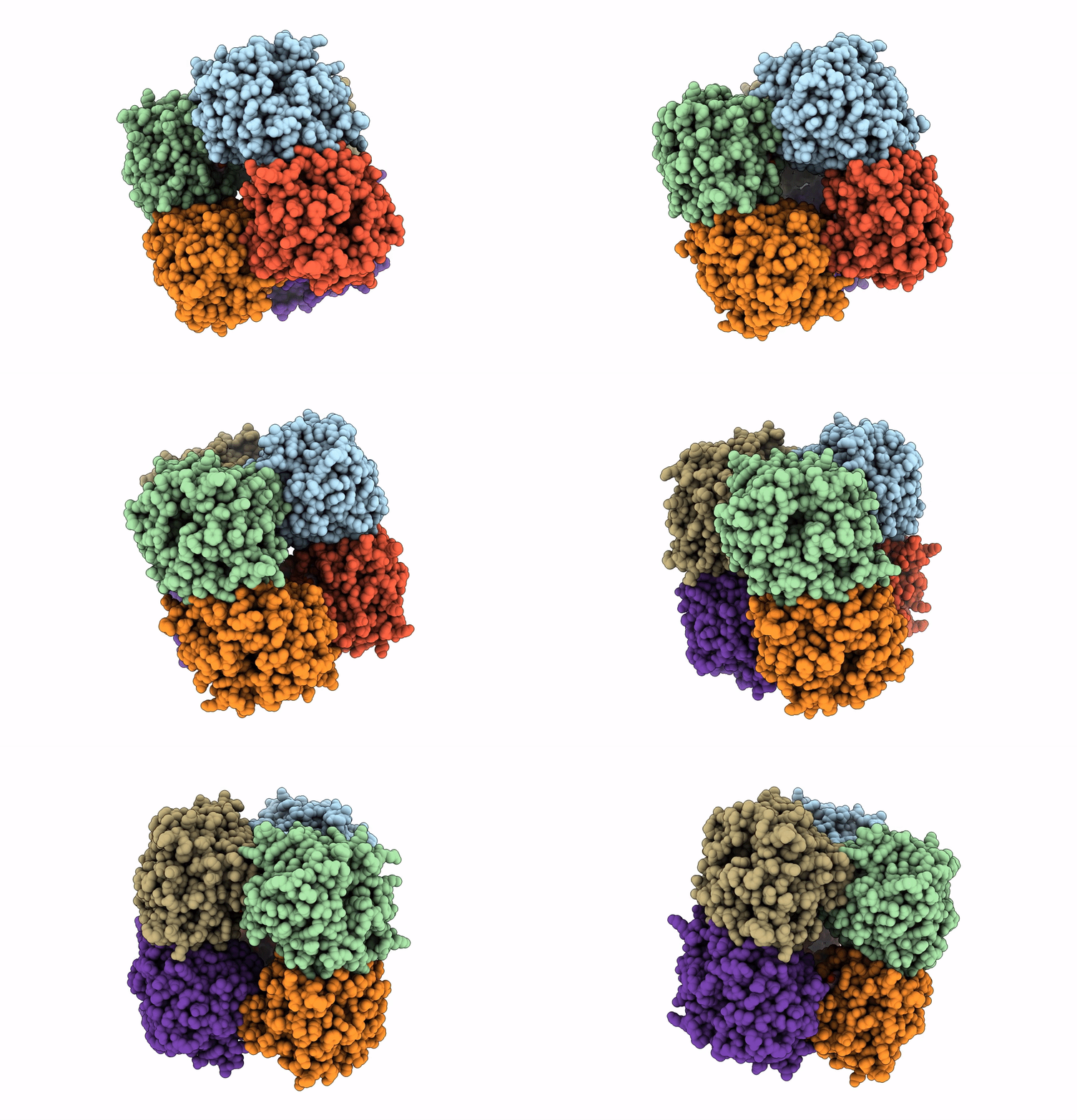
5AYE image
Entry Detail
PDB ID:
5AYE
Keywords:
Title:
Crystal structure of Ruminococcus albus beta-(1,4)-mannooligosaccharide phosphorylase (RaMP2) in complexes with phosphate and beta-(1,4)-mannobiose
Biological Source:
PDB Version:
Deposition Date:
2015-08-13
Release Date:
2016-03-23
Method Details:
Experimental Method:
Resolution:
2.20 Å
R-Value Free:
0.21
R-Value Work:
0.16
R-Value Observed:
0.16
Space Group:
P 1 21 1
Macromolecular Entities
Polymer Type:polypeptide(L)
Description:Beta-1,4-mannooligosaccharide phosphorylase
Chain IDs:A, B, C, D, E, F
Chain Length:335
Number of Molecules:6
Biological Source:Ruminococcus albus (strain ATCC 27210 / DSM 20455 / JCM 14654 / NCDO 2250 / 7)
Ligand Molecules
Peptide-like Molecules
PRD_900115
Primary Citation
Structural insights into the difference in substrate recognition of two mannoside phosphorylases from two GH130 subfamilies.
Febs Lett. 590 828 837 (2016)
PMID: 26913570 DOI: 10.1002/1873-3468.12105

Abstact

In Ruminococcus albus, 4-O-β-D-mannosyl-D-glucose phosphorylase (RaMP1) and β-(1,4)-mannooligosaccharide phosphorylase (RaMP2) belong to two subfamilies of glycoside hydrolase family 130. The two enzymes phosphorolyze β-mannosidic linkages at the nonreducing ends of their substrates, and have substantially diverse substrate specificity. The differences in their mechanism of substrate binding have not yet been fully clarified. In the present study, we report the crystal structures of RaMP1 with/without 4-O-β-D-mannosyl-d-glucose and RaMP2 with/without β-(1→4)-mannobiose. The structures of the two enzymes differ at the +1 subsite of the substrate-binding pocket. Three loops are proposed to determine the different substrate specificities. One of these loops is contributed from the adjacent molecule of the oligomer structure. In RaMP1, His245 of loop 3 forms a hydrogen-bond network with the substrate through a water molecule, and is indispensible for substrate binding.

Legend

Protein

Chemical

Disease

Primary Citation of related structures