
Deposition Date
2015-08-11
Release Date
2016-03-23
Last Version Date
2024-03-20
Entry Detail
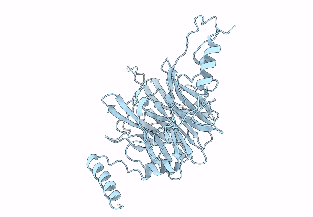
PDB ID:
5AY9
Keywords:
Title:
Crystal structure of Ruminococcus albus 4-O-beta-D-mannosyl-D-glucose phosphorylase (RaMP1)
Biological Source:
Source Organism(s):
Expression System(s):
Method Details:
Experimental Method:
Resolution:
2.50 Å
R-Value Free:
0.28
R-Value Work:
0.23
R-Value Observed:
0.24
Space Group:
H 3 2
