
Deposition Date
2015-07-29
Release Date
2016-03-02
Last Version Date
2024-11-13
Entry Detail
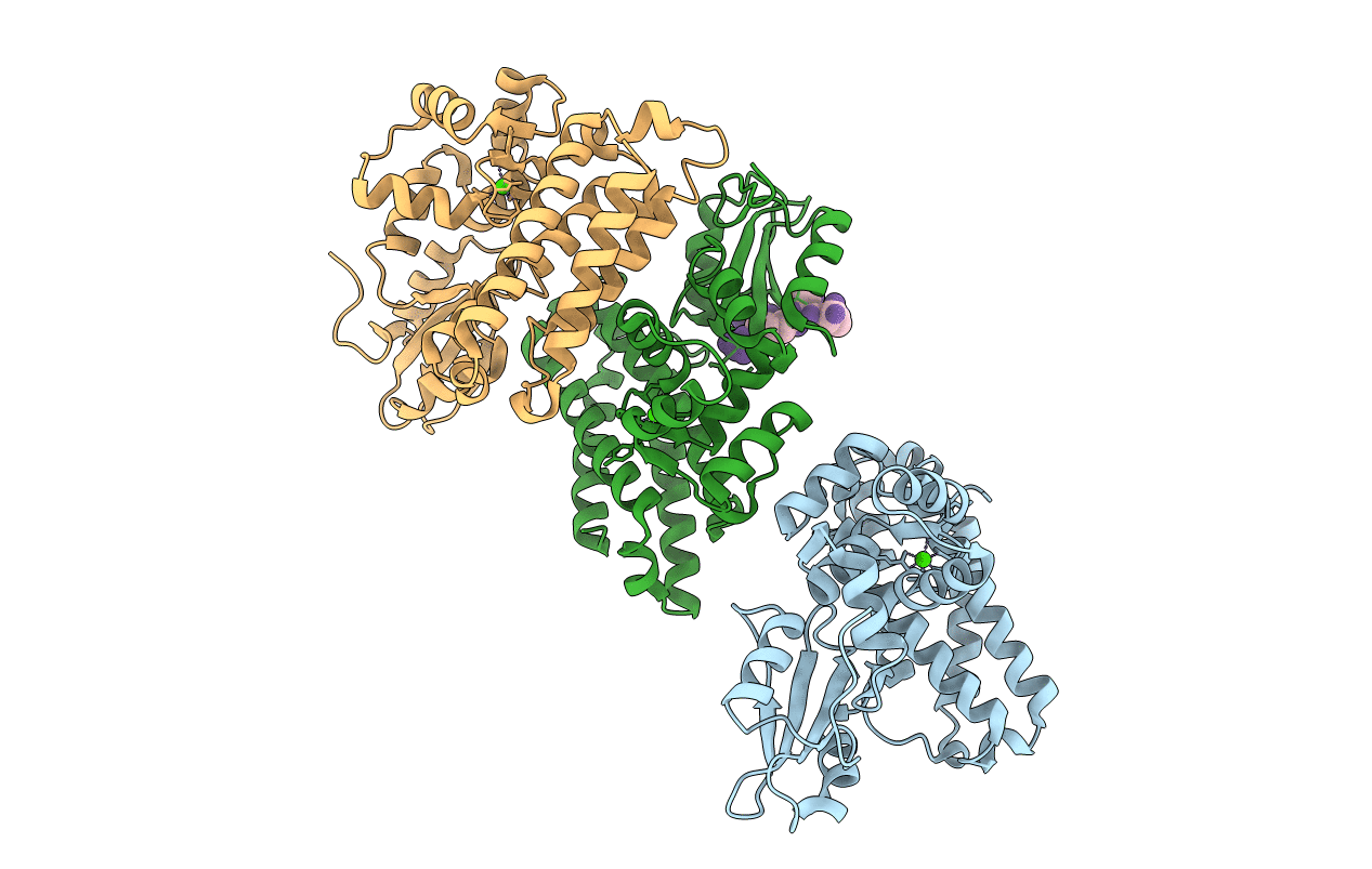
PDB ID:
5AXI
Keywords:
Title:
Crystal structure of Cbl-b TKB domain in complex with Cblin
Biological Source:
Source Organism(s):
Mus musculus (Taxon ID: 10090)
Expression System(s):
Method Details:
Experimental Method:
Resolution:
2.50 Å
R-Value Free:
0.26
R-Value Work:
0.19
R-Value Observed:
0.20
Space Group:
P 21 21 21
