5AWM image
Deposition Date 2015-07-06
Release Date 2015-08-05
Last Version Date 2025-03-19
Entry Detail
PDB ID:
5AWM
Keywords:
Title:
The Crystal Structure of JNK from Drosophila melanogaster Reveals an Evolutionarily Conserved Topology with that of Mammalian JNK Proteins.
Biological Source:
Source Organism:
Host Organism:
Method Details:
Experimental Method:
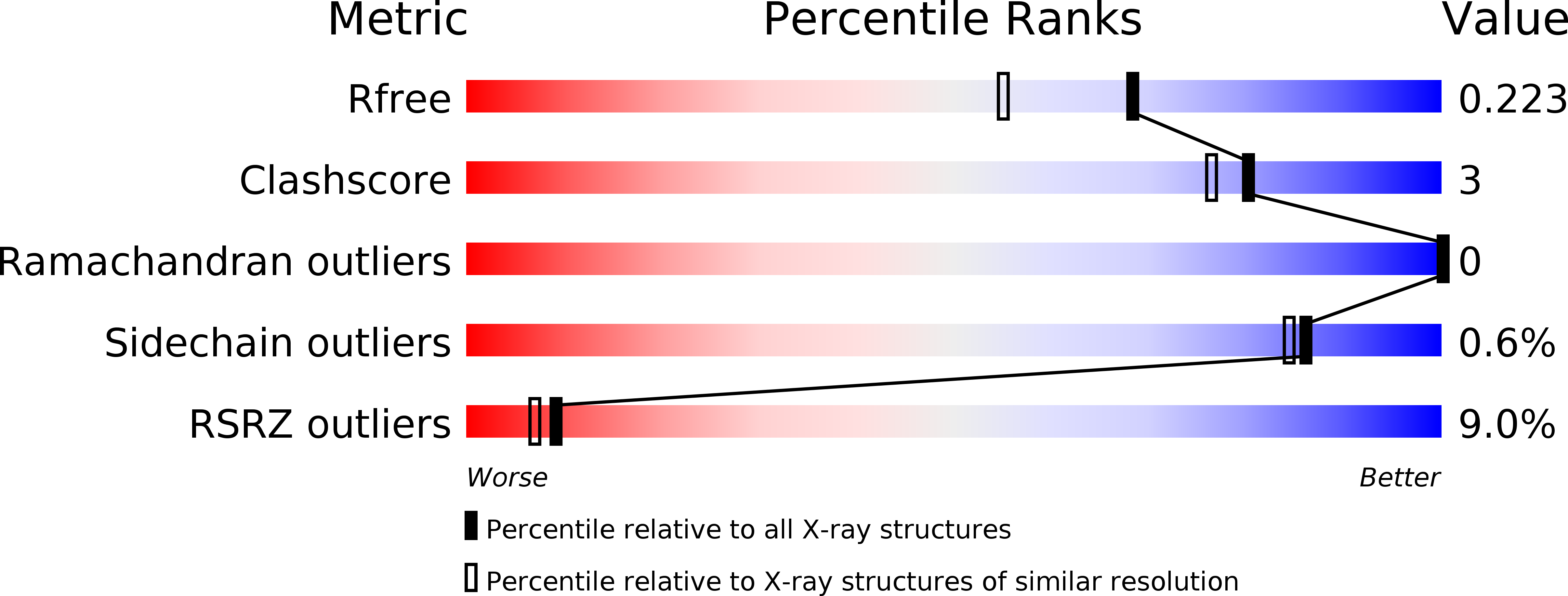
Resolution:
1.79 Å
R-Value Free:
0.21
R-Value Work:
0.17
R-Value Observed:
0.17
Space Group:
P 21 21 21
Macromolecular Entities
Structures with similar UniProt ID
Protein Blast
Polymer Type:polypeptide(L)
Molecule:Stress-activated protein kinase JNK
Gene (Uniprot):bsk
Chain IDs:A
Chain Length:394
Number of Molecules:1
Biological Source:Drosophila melanogaster
Primary Citation
The crystal structure of JNK from Drosophila melanogaster reveals an evolutionarily conserved topology with that of mammalian JNK proteins.
Bmc Struct.Biol. 15 17 17 (2015)
PMID: 26377800 DOI: 10.1186/s12900-015-0045-1

Abstact

BACKGROUND The c-Jun N-terminal kinases (JNKs), members of the mitogen-activated protein kinase (MAPK) family, engage in diverse cellular responses to signals produced under normal development and stress conditions. In Drosophila, only one JNK member is present, whereas ten isoforms from three JNK genes (JNK1, 2, and 3) are present in mammalian cells. To date, several mammalian JNK structures have been determined, however, there has been no report of any insect JNK structure. RESULTS We report the first structure of JNK from Drosophila melanogaster (DJNK). The crystal structure of the unphosphorylated form of DJNK complexed with adenylyl imidodiphosphate (AMP-PNP) has been solved at 1.79 Å resolution. The fold and topology of DJNK are similar to those of mammalian JNK isoforms, demonstrating their evolutionarily conserved structures and functions. Structural comparisons of DJNK and the closely related mammalian JNKs also allow identification of putative catalytic residues, substrate-binding sites and conformational alterations upon docking interaction with Drosophila scaffold proteins. CONCLUSIONS The DJNK structure reveals common features with those of the mammalian JNK isoforms, thereby allowing the mapping of putative catalytic and substrate binding sites. Additionally, structural changes upon peptide binding could be predicted based on the comparison with the closely-related JNK3 structure in complex with pepJIP1. This is the first structure of insect JNK reported to date, and will provide a platform for future mutational studies in Drosophila to ascertain the functional role of insect JNK.

Legend

Protein

Chemical

Disease

Primary Citation of related structures