Deposition Date
2015-07-01
Release Date
2015-09-02
Last Version Date
2024-11-13
Entry Detail
PDB ID:
5AW9
Keywords:
Title:
Kinetics by X-ray crystallography: native E2.MgF42-.2K+ crystal for Rb+ bound crystals
Biological Source:
Source Organism(s):
Squalus acanthias (Taxon ID: 7797)
Method Details:
Experimental Method:
Resolution:
2.80 Å
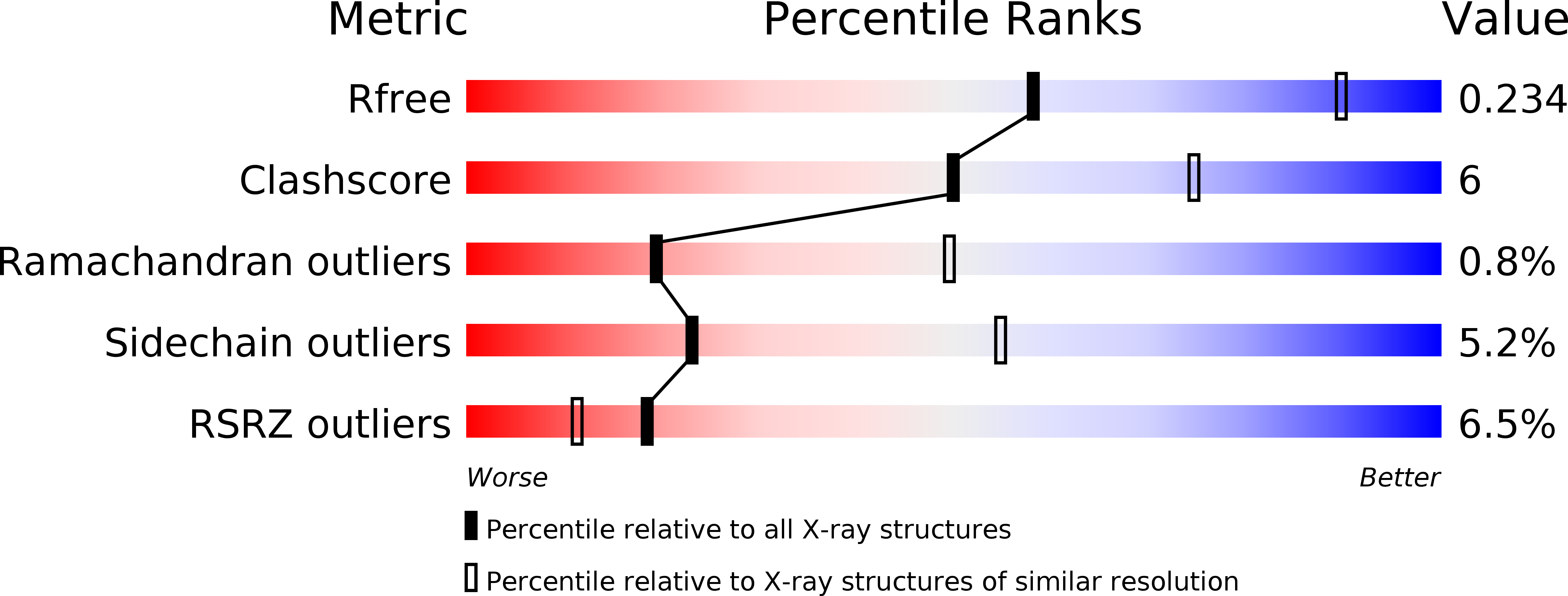
R-Value Free:
0.26
R-Value Work:
0.21
R-Value Observed:
0.21
Space Group:
C 1 2 1