5AVM image
Entry Detail
PDB ID:
5AVM
Keywords:
Title:
Crystal structures of 5-aminoimidazole ribonucleotide (AIR) synthetase, PurM, from Thermus thermophilus
Biological Source:
Source Organism:
Host Organism:
PDB Version:
Deposition Date:
2015-06-23
Release Date:
2015-11-25
Method Details:
Experimental Method:
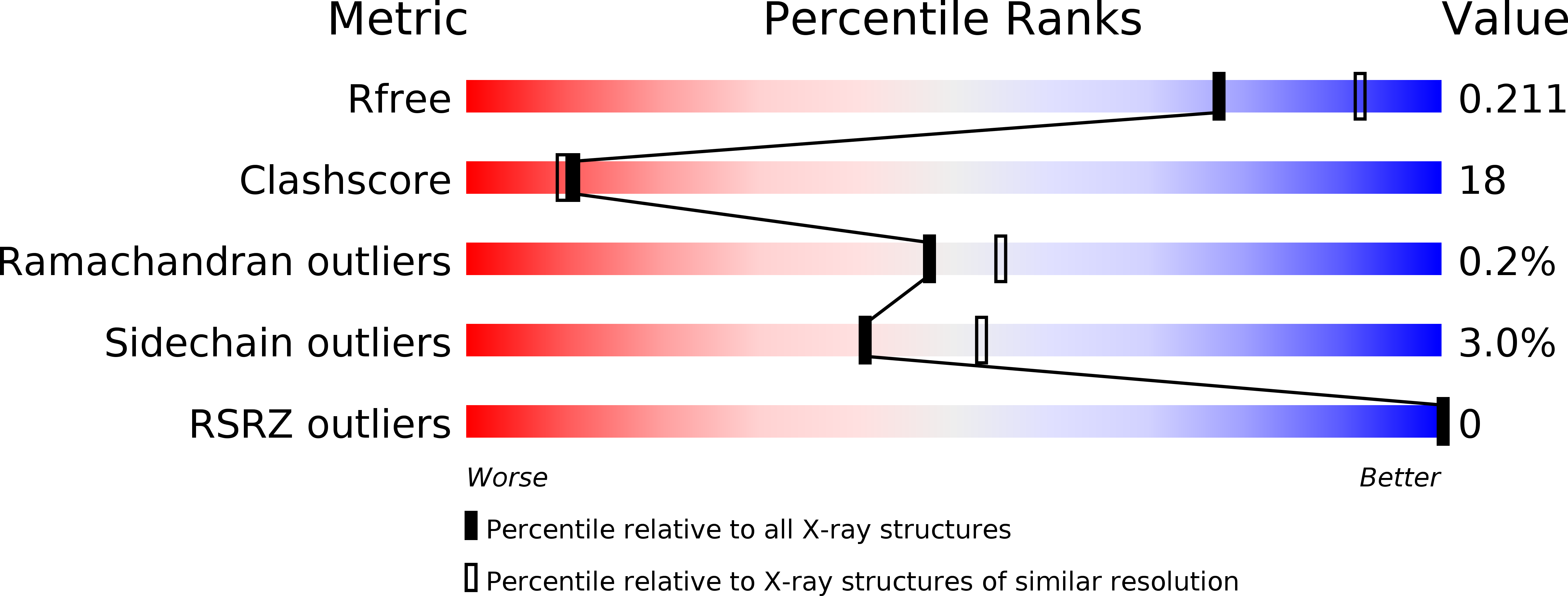
Resolution:
2.20 Å
R-Value Free:
0.21
R-Value Work:
0.16
R-Value Observed:
0.16
Space Group:
P 21 21 2
Macromolecular Entities
Polymer Type:polypeptide(L)
Description:Phosphoribosylformylglycinamidine cyclo-ligase
Chain IDs:A, B, C, D, E, F, G, H
Chain Length:333
Number of Molecules:8
Biological Source:Thermus thermophilus HB8
Ligand Molecules
Primary Citation
Crystal structures and ligand binding of PurM proteins from Thermus thermophilus and Geobacillus kaustophilus
J.Biochem. 159 313 321 (2016)
PMID: 26515187 DOI: 10.1093/jb/mvv107

Abstact

Crystal structures of 5-aminoimidazole ribonucleotide (AIR) synthetase, also known as PurM, from Thermus thermophilus (Tt) and Geobacillus kaustophilus (Gk) were determined. For TtPurM, the maximum resolution was 2.2 Å and the space group was P21212 with four dimers in an asymmetric unit. For GkPurM, the maximum resolution was 2.2 Å and the space group was P21212 with one monomer in asymmetric unit. The biological unit is dimer for both TtPurM and GkPurM and the dimer structures were similar to previously determined structures of PurM in general. For TtPurM, ∼50 residues at the amino terminal were disordered in the crystal structure whereas, for GkPurM, the corresponding region covered the ATP-binding site forming an α helix in part, suggesting that the N-terminal region of PurM changes its conformation upon binding of ligands. FGAM binding site was predicted by the docking simulation followed by the MD simulation based on the SO4 (2-) binding site found in the crystal structure of TtPurM.

Legend

Protein

Chemical

Disease

Primary Citation of related structures