5ARL image
Deposition Date 2015-09-25
Release Date 2016-01-20
Last Version Date 2024-11-13
Entry Detail
PDB ID:
5ARL
Keywords:
Title:
crystal structure of porcine RNase 4 D80A mutant in complex with dCMP
Biological Source:
Source Organism:
SUS SCROFA (Taxon ID: 9823)
Host Organism:
Method Details:
Experimental Method:
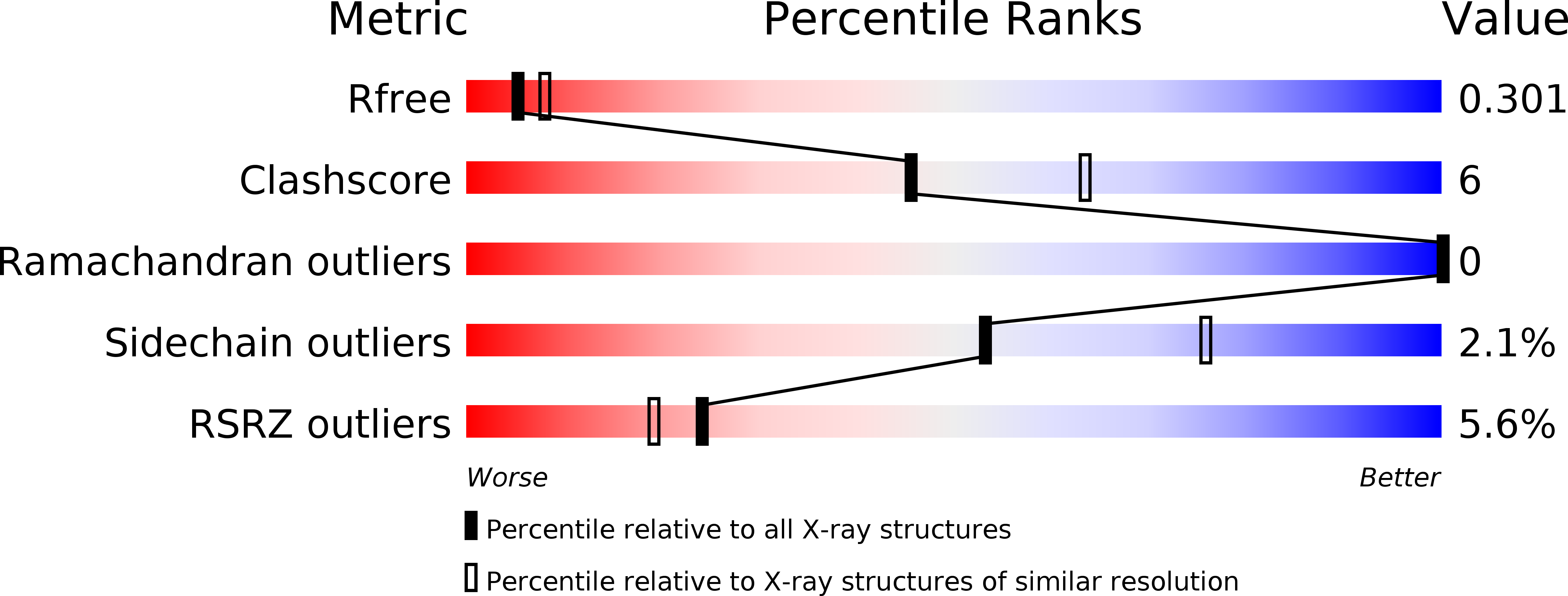
Resolution:
2.61 Å
R-Value Free:
0.29
R-Value Work:
0.25
R-Value Observed:
0.25
Space Group:
P 1
Macromolecular Entities
Polymer Type:polypeptide(L)
Molecule:RIBONUCLEASE 4
Gene (Uniprot):RNASE4
Mutations:YES
Chain IDs:A, B, C, D
Chain Length:134
Number of Molecules:4
Biological Source:SUS SCROFA
Ligand Molecules
Primary Citation
Structural Basis of Substrate Specificity in Porcine Rnase 4.
FEBS J. 283 912 ? (2016)
PMID: 26748441 DOI: 10.1111/FEBS.13646

Abstact

UNLABELLED RNase 4, a member of the RNase A superfamily with substrate preference for uridine, has roles in host defence, angiogenesis and neurodegenerative diseases. It also exhibits the highest interspecies amino acid sequence similarity amongst RNase A family members. However, compared to other members of the RNase A family, including eosinophil-derived neurotoxin, eosinophil cationic protein and angiogenin, little is known about the molecular basis of substrate specificity in RNase 4. Here we report high to medium resolution structures of native porcine RNase 4 (PL3), a 'substrate-specificity' determining mutant D80A and their respective complexes with deoxyuridine 5'-monophosphate (dUMP) and deoxycytidine 5'-monophosphate (dCMP). These structures provide insight into the structural basis of the uridine versus cytosine substrate specificity in RNase 4: in the D80A mutant (D80A•dCMP), the side chain of Arg101 is positioned further away from the substrate-binding pocket due to the loss of the Asp80 side chain, reducing the repulsion force on the less favoured dCMP from Arg101 and allowing the ligand to occupy the binding pocket. This can also explain the observation that the ligand in the D80A•dCMP complex is stabilized only by a small number of hydrogen bonds. Compared to the previously reported structure of the human RNase 4•2'-deoxyuridine 3'-phosphate complex, the structure of PL3•dUMP complex shows additional hydrogen bonds between the ligand and the protein. In addition, the interaction between Arg101 and the dUMP ligand is absent. These observed differences are probably the result of the flexibility and different 'positioning' of the phosphate group among the mononucleotide ligands. DATABASE The atomic coordinates and structure factors for PL3 (5AR6), D80A (5ARJ), PL3∙dUMP (5ARK) and D80A∙dCMP (5ARL) complexes have been deposited with the Protein Data Bank, Research Collaboratory for Structural Bioinformatics, Rutgers University, New Brunswick, NJ, USA (http://www.rcsb.org/).

Legend

Protein

Chemical

Disease

Primary Citation of related structures