
Deposition Date
2015-09-17
Release Date
2016-01-27
Last Version Date
2024-01-10
Entry Detail
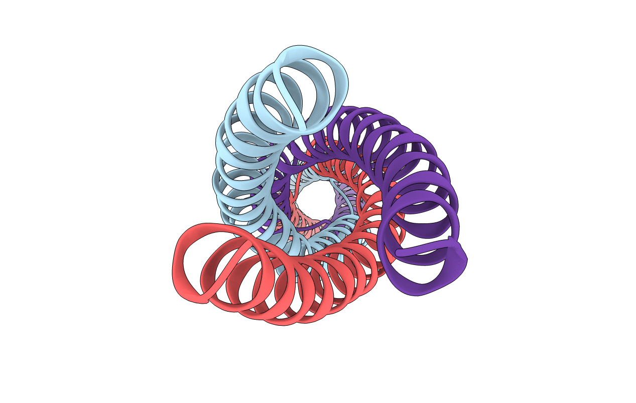
PDB ID:
5APP
Keywords:
Title:
Actinobacillus actinomycetemcomitans OMP100 residues 133-198 fused to GCN4 adaptors
Biological Source:
Source Organism(s):
Expression System(s):
Method Details:
Experimental Method:
Resolution:
2.30 Å
R-Value Free:
0.25
R-Value Work:
0.22
R-Value Observed:
0.22
Space Group:
C 1 2 1
