5AMK image
Entry Detail
PDB ID:
5AMK
Title:
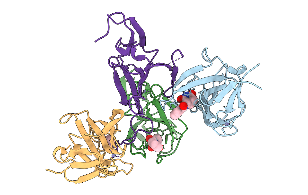
Cereblon isoform 4 from Magnetospirillum gryphiswaldense in multiple conformations, hexagonal crystal form
Biological Source:
Host Organism:
PDB Version:
Deposition Date:
2015-03-10
Release Date:
2015-07-01
Method Details:
Experimental Method:
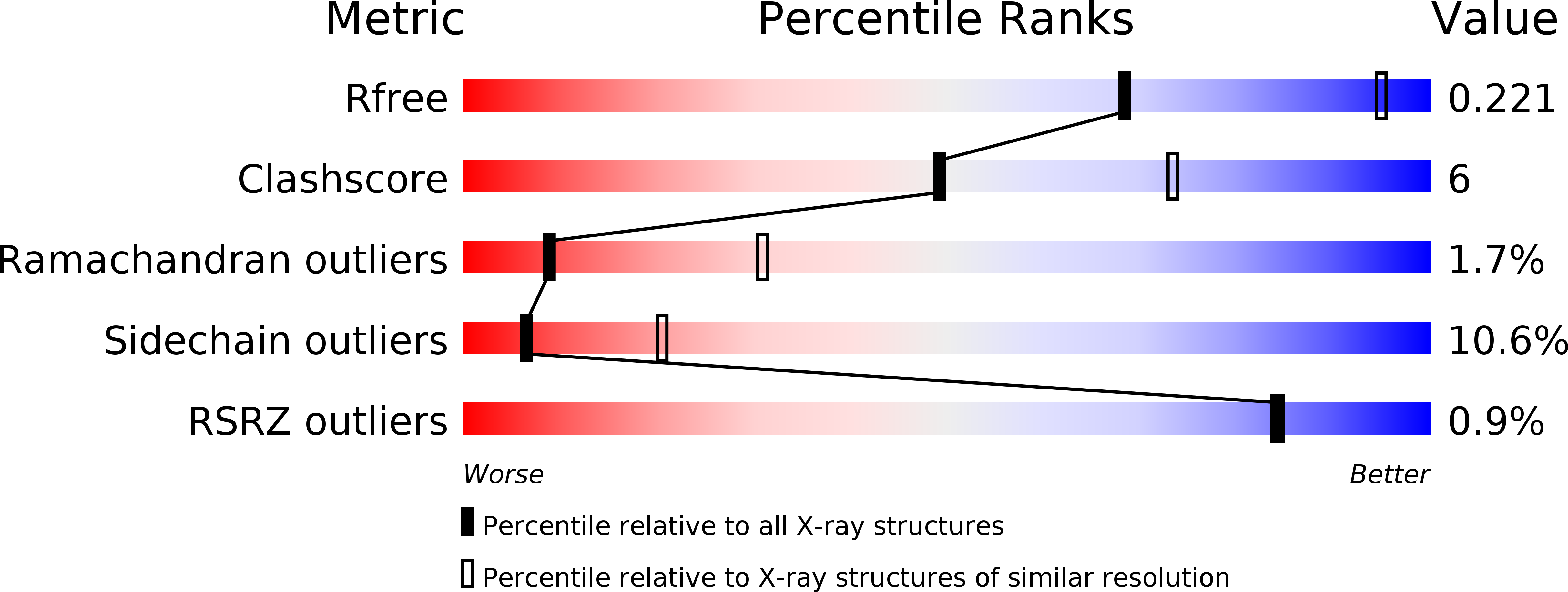
Resolution:
2.90 Å
R-Value Free:
0.21
R-Value Work:
0.17
R-Value Observed:
0.17
Space Group:
P 61 2 2
Macromolecular Entities
Polymer Type:polypeptide(L)
Description:CEREBLON ISOFORM 4
Chain IDs:A, B, C, D
Chain Length:125
Number of Molecules:4
Biological Source:MAGNETOSPIRILLUM GRYPHISWALDENSE
Primary Citation
Structural Dynamics of the Cereblon Ligand Binding Domain.
Plos One 10 28342 ? (2015)
PMID: 26024445 DOI: 10.1371/JOURNAL.PONE.0128342

Abstact

Cereblon, a primary target of thalidomide and its derivatives, has been characterized structurally from both bacteria and animals. Especially well studied is the thalidomide binding domain, CULT, which shows an invariable structure across different organisms and in complex with different ligands. Here, based on a series of crystal structures of a bacterial representative, we reveal the conformational flexibility and structural dynamics of this domain. In particular, we follow the unfolding of large fractions of the domain upon release of thalidomide in the crystalline state. Our results imply that a third of the domain, including the thalidomide binding pocket, only folds upon ligand binding. We further characterize the structural effect of the C-terminal truncation resulting from the mental-retardation linked R419X nonsense mutation in vitro and offer a mechanistic hypothesis for its irresponsiveness to thalidomide. At 1.2Å resolution, our data provide a view of thalidomide binding at atomic resolution.

Legend

Protein

Chemical

Disease

Primary Citation of related structures