Deposition Date
2015-03-02
Release Date
2015-07-22
Last Version Date
2024-05-08
Entry Detail
PDB ID:
5AK8
Keywords:
Title:
Structure of C351A mutant of Porphyromonas gingivalis peptidylarginine deiminase
Biological Source:
Source Organism(s):
PORPHYROMONAS GINGIVALIS (Taxon ID: 837)
Expression System(s):
Method Details:
Experimental Method:
Resolution:
1.48 Å
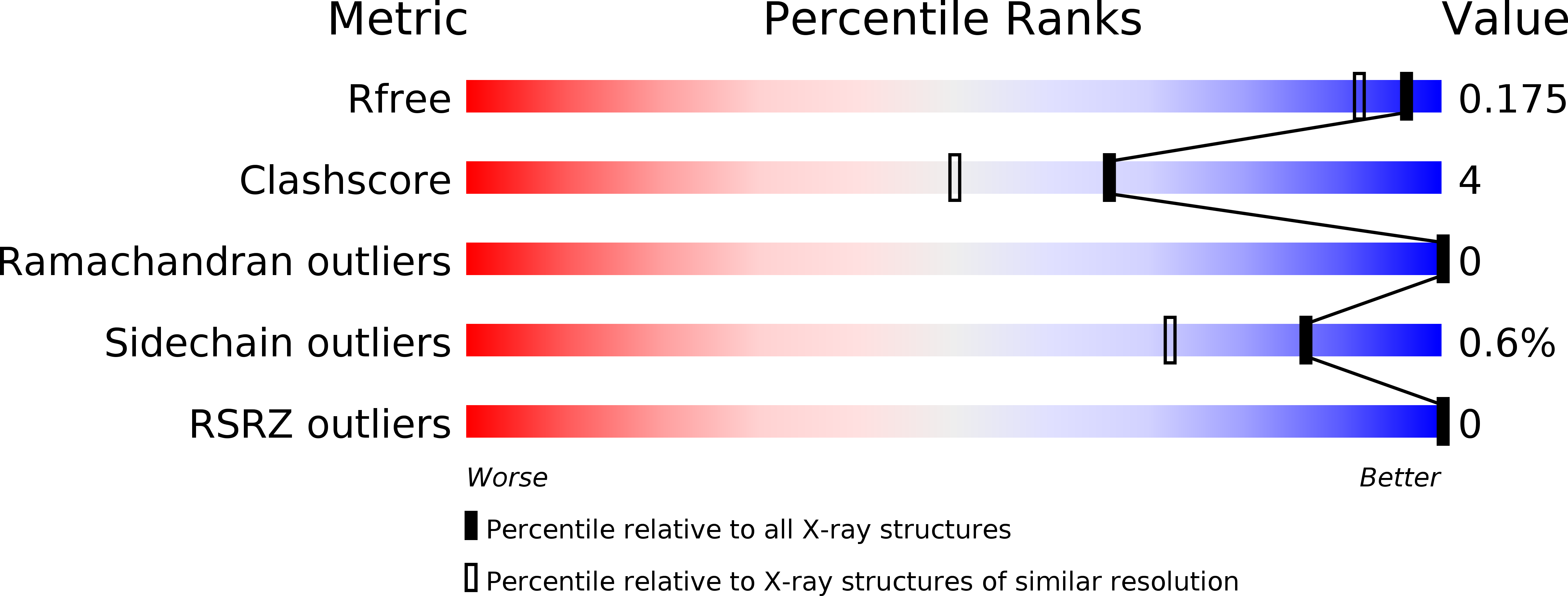
R-Value Free:
0.16
R-Value Work:
0.12
R-Value Observed:
0.12
Space Group:
C 1 2 1