
Deposition Date
2015-02-24
Release Date
2016-03-09
Last Version Date
2024-11-20
Entry Detail
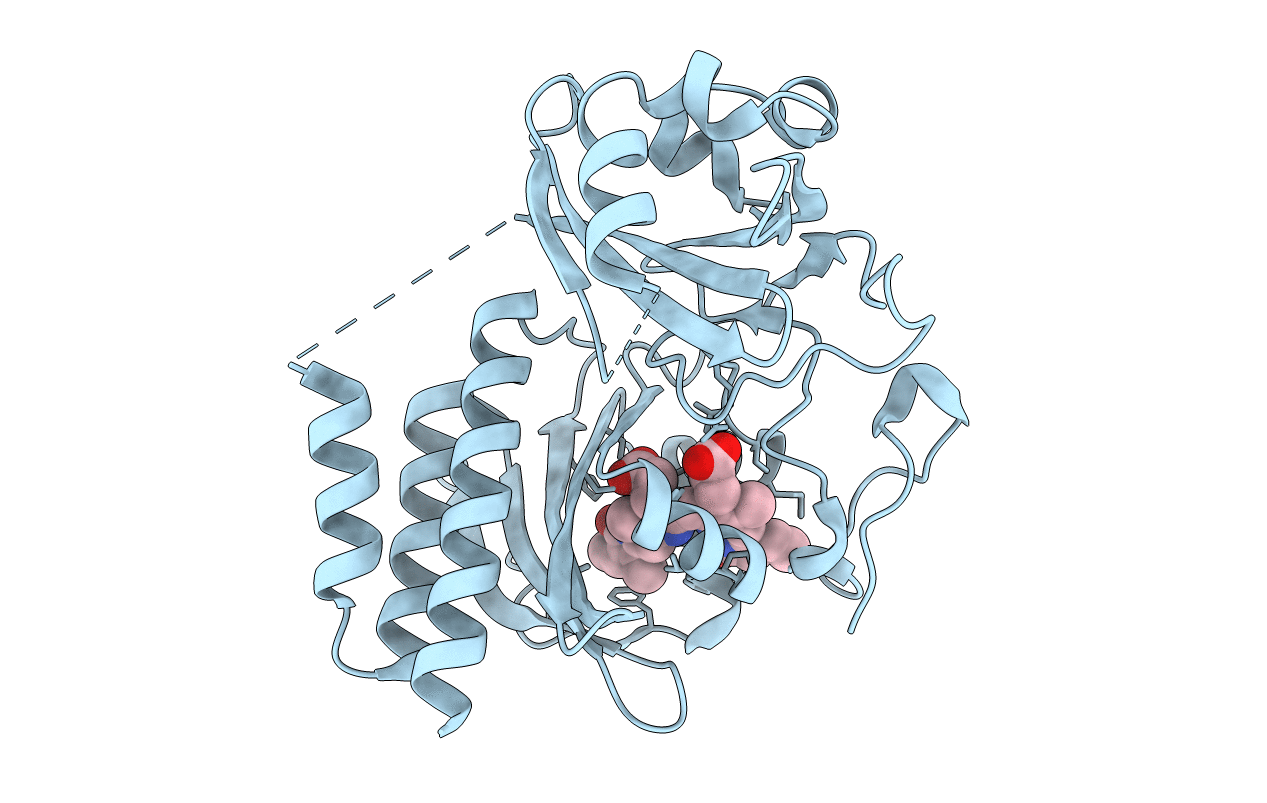
PDB ID:
5AJG
Keywords:
Title:
Structure of Infrared Fluorescent Protein IFP1.4 AT 1.11 Angstrom resolution
Biological Source:
Source Organism:
DEINOCOCCUS RADIODURANS (Taxon ID: 1299)
Host Organism:
Method Details:
Experimental Method:
Resolution:
1.11 Å
R-Value Free:
0.16
R-Value Work:
0.14
R-Value Observed:
0.14
Space Group:
C 1 2 1
