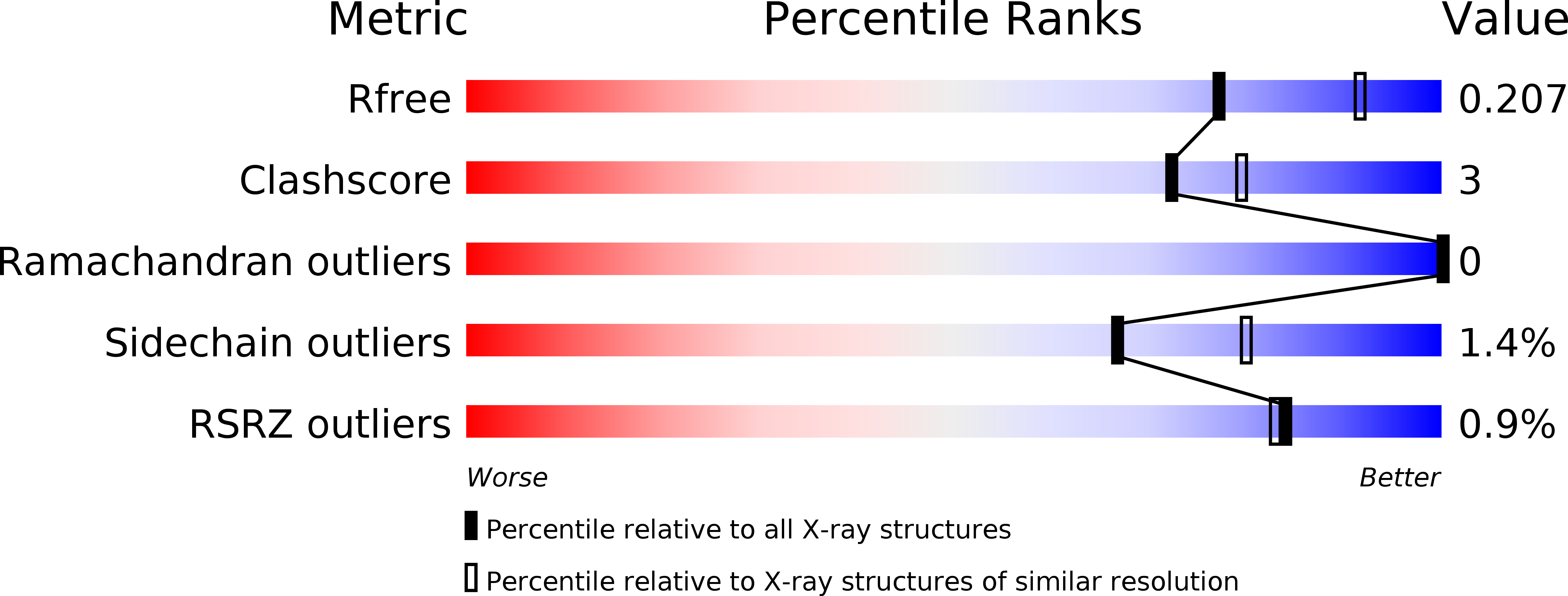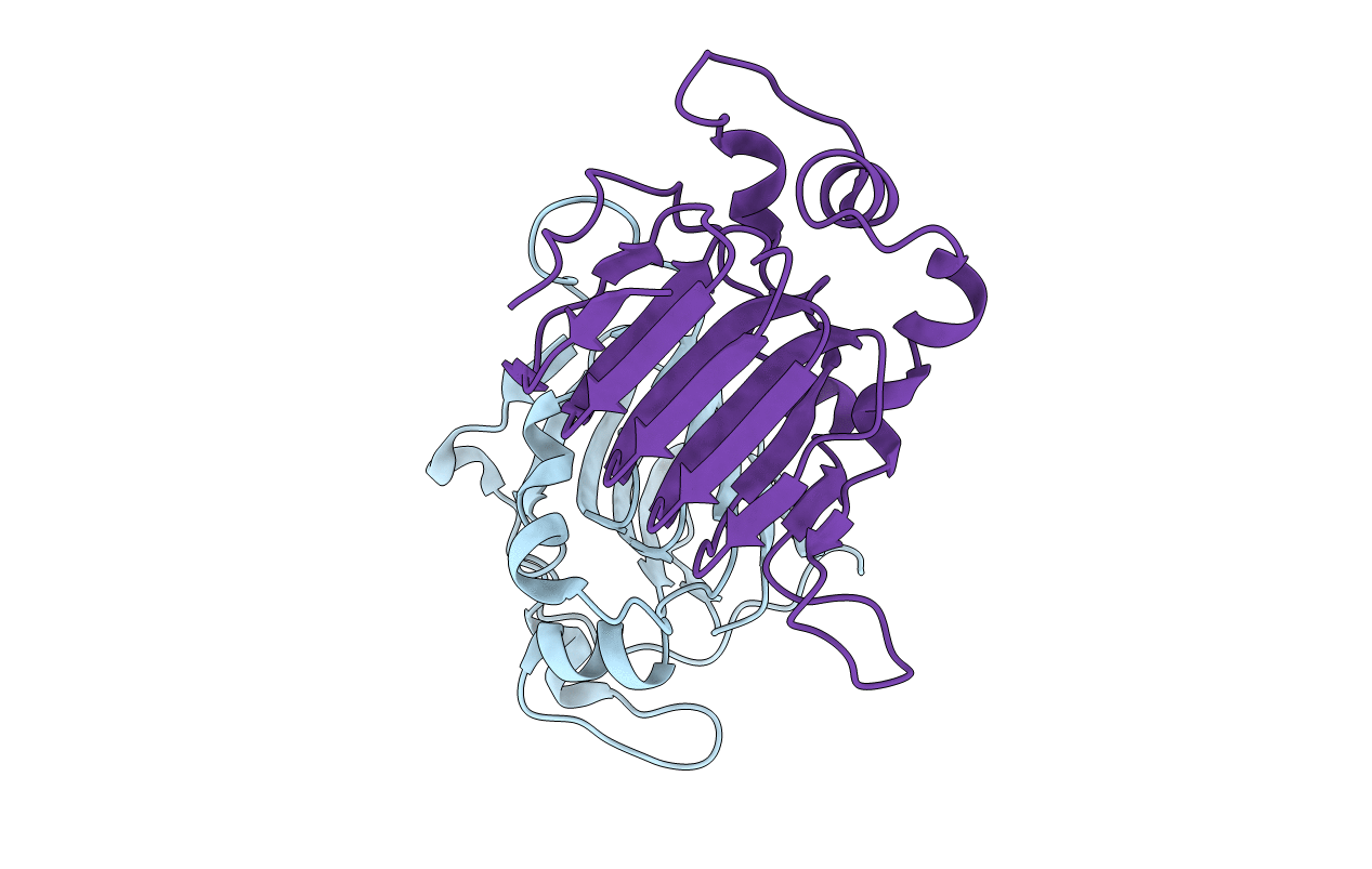
5AJ8 image
Entry Detail
PDB ID:
5AJ8
Title:
Tubulin Binding Cofactor C from Leishmania major
Biological Source:
Source Organism:
Host Organism:
PDB Version:
Deposition Date:
2015-02-20
Release Date:
2015-04-15
Method Details:
Experimental Method:
Resolution:
2.20 Å
R-Value Free:
0.19
R-Value Work:
0.15
R-Value Observed:
0.15
Space Group:
P 1 21 1
Macromolecular Entities
Polymer Type:polypeptide(L)
Description:TUBULIN BINDING COFACTOR C
Mutations:YES
Chain IDs:A, B
Chain Length:187
Number of Molecules:2
Biological Source:LEISHMANIA MAJOR
Modified Residue
Compound ID Chain ID Parent Comp ID Details 2D Image
MSE A MET SELENOMETHIONINE
Primary Citation
Crystal Structure of the C-Terminal Domain of Tubulin-Binding Cofactor C from Leishmania Major.
Mol.Biochem.Parasitol. 201 26 ? (2015)
PMID: 25982270 DOI: 10.1016/J.MOLBIOPARA.2015.05.003

Abstact

Tubulin-binding cofactor C stimulates GTPase activity and contributes to the release of the heterodimeric α/β-tubulin from a super-complex of tubulin monomers and two ancillary cofactors. We have determined the 2.2 Å resolution crystal structure of the C-terminal domain of tubulin-binding cofactor C from Leishmania major based on single wavelength anomalous dispersion measurements targeting a selenomethionine derivative. Although previously predicted to consist of two domains the structure is best described as a single domain dominated by a right-handed β-helix of five turns that form a triangular prism. One face of the prism is covered by the C-terminal residues leaving another face solvent exposed. Comparisons with an orthologous human GTPase activating protein match key residues involved in binding nucleotide and identify the face of the β-helix fold likely involved in interacting with the β-tubulin:GTP complex.

Legend

Protein

Chemical

Disease

Primary Citation of related structures