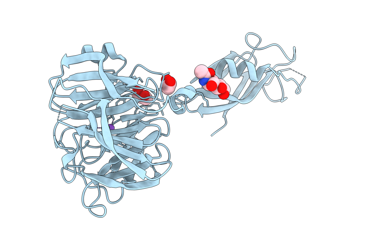
5AFB image
Entry Detail
PDB ID:
5AFB
Title:
Crystal structure of the Latrophilin3 Lectin and Olfactomedin Domains
Biological Source:
Source Organism:
Host Organism:
PDB Version:
Deposition Date:
2015-01-21
Release Date:
2015-03-18
Method Details:
Experimental Method:
Resolution:
2.16 Å
R-Value Free:
0.24
R-Value Work:
0.20
R-Value Observed:
0.20
Space Group:
I 2 2 2
Macromolecular Entities
Polymer Type:polypeptide(L)
Description:LATROPHILIN-3
Chain IDs:A
Chain Length:384
Number of Molecules:1
Biological Source:HOMO SAPIENS
Modified Residue
Compound ID Chain ID Parent Comp ID Details 2D Image
ASN A ASN GLYCOSYLATION SITE
Primary Citation
Structural Basis of Latrophilin-Flrt Interaction.
Structure 23 774 ? (2015)
PMID: 25728924 DOI: 10.1016/J.STR.2015.01.013

Abstact

Latrophilins, receptors for spider venom α-latrotoxin, are adhesion type G-protein-coupled receptors with emerging functions in synapse development. The N-terminal region binds the endogenous cell adhesion molecule FLRT, a major regulator of cortical and synapse development. We present crystallographic data for the mouse Latrophilin3 lectin and olfactomedin-like (Olf) domains, thereby revealing the Olf β-propeller fold and conserved calcium-binding site. We locate the FLRT-Latrophilin binding surfaces by a combination of sequence conservation analysis, point mutagenesis, and surface plasmon resonance experiments. In stripe assays, we show that wild-type Latrophilin3 and its high-affinity interactor FLRT2, but not the binding-impaired mutants we generated, promote HeLa cell adhesion. In contrast, cortical neurons expressing endogenous FLRTs are repelled by wild-type Latrophilin3 and not by the binding-impaired mutant. Taken together, we present molecular level insights into Latrophilin structure, its FLRT-binding mechanism, and a role for Latrophilin and FLRT that goes beyond a simply adhesive interaction.

Legend

Protein

Chemical

Disease

Primary Citation of related structures