5AES image
Deposition Date 2015-01-09
Release Date 2015-04-01
Last Version Date 2024-10-23
Entry Detail
PDB ID:
5AES
Keywords:
Title:
Crystal Structure of murine Chronophin (Pyridoxal Phosphate Phosphatase) in Complex with a PNP-derived Inhibitor
Biological Source:
Source Organism:
MUS MUSCULUS (Taxon ID: 10090)
Host Organism:
Method Details:
Experimental Method:
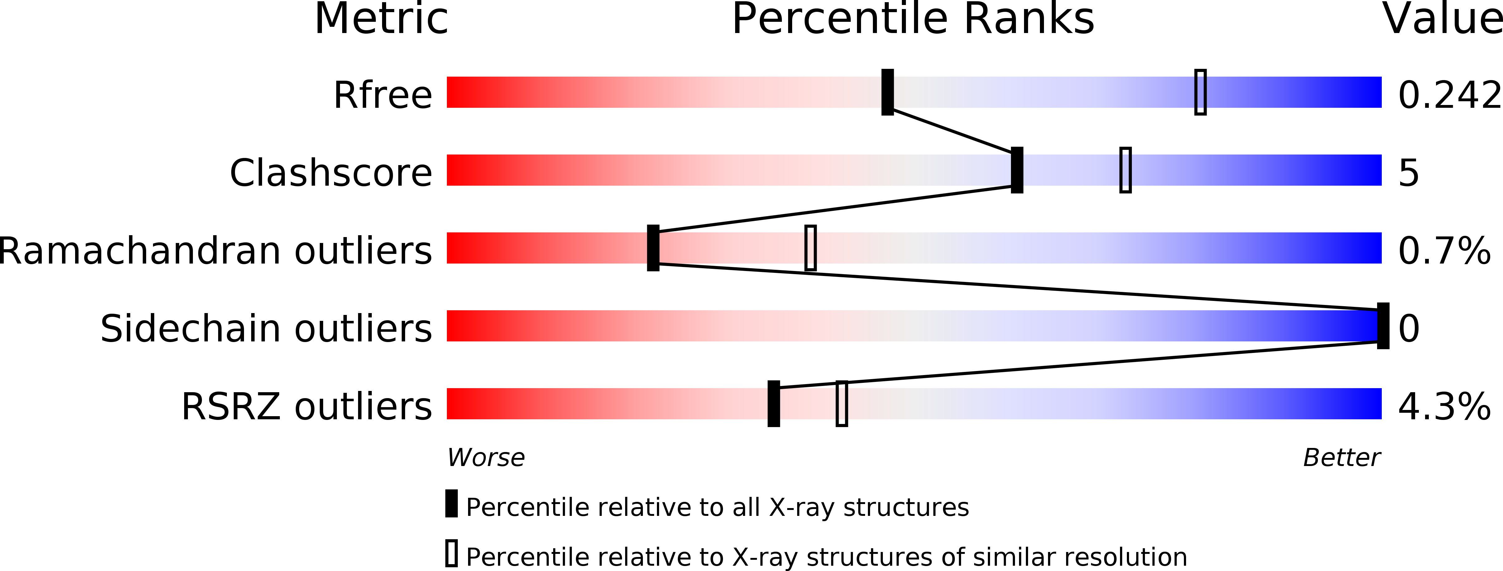
Resolution:
2.75 Å
R-Value Free:
0.24
R-Value Work:
0.18
R-Value Observed:
0.19
Space Group:
I 2 3
Macromolecular Entities
Polymer Type:polypeptide(L)
Molecule:PYRIDOXAL PHOSPHATE PHOSPHATASE
Gene (Uniprot):Pdxp
Chain IDs:A, B
Chain Length:293
Number of Molecules:2
Biological Source:MUS MUSCULUS
Primary Citation
Synthesis of Hydrolysis-Resistant Pyridoxal 5'-Phosphate Analogs and Their Biochemical and X-Ray Crystallographic Characterization with the Pyridoxal Phosphatase Chronophin.
Bioorg.Med.Chem. 23 2819 ? (2015)
PMID: 25783190 DOI: 10.1016/J.BMC.2015.02.049

Abstact

A set of phosphonic acid derivatives (1-4) of pyridoxal 5'-phosphate (PLP) was synthesized and characterized biochemically using purified murine pyridoxal phosphatase (PDXP), also known as chronophin. The most promising compound 1 displayed primarily competitive PDXP inhibitory activity with an IC50 value of 79μM, which was in the range of the Km of the physiological substrate PLP. We also report the X-ray crystal structure of PDXP bound to compound 3, which we solved to 2.75Å resolution (PDB code 5AES). The co-crystal structure proves that compound 3 binds in the same orientation as PLP, and confirms the mode of inhibition to be competitive. Thus, we identify compound 1 as a PDXP phosphatase inhibitor. Our results suggest a strategy to design new, potent and selective PDXP inhibitors, which may be useful to increase the sensitivity of tumor cells to treatment with cytotoxic agents.

Legend

Protein

Chemical

Disease

Primary Citation of related structures