5AEC image
Entry Detail
PDB ID:
5AEC
Keywords:
Title:
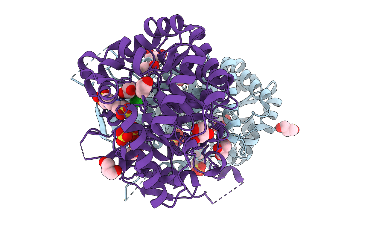
Type II Baeyer-Villiger monooxygenase.The oxygenating constituent of 3,6-diketocamphane monooxygenase from CAM plasmid of Pseudomonas putida in complex with FMN.
Biological Source:
Source Organism:
Host Organism:
PDB Version:
Deposition Date:
2015-08-28
Release Date:
2015-09-09
Method Details:
Experimental Method:
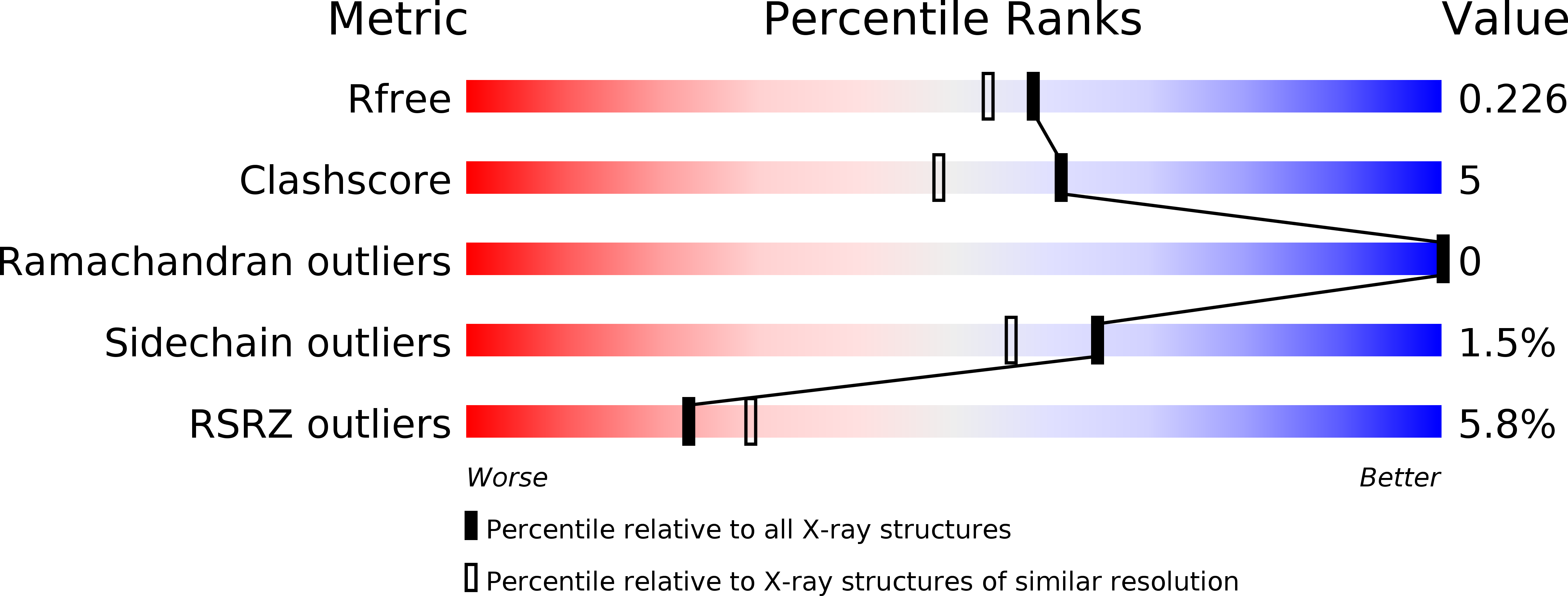
Resolution:
1.93 Å
R-Value Free:
0.22
R-Value Work:
0.17
R-Value Observed:
0.17
Space Group:
P 21 21 21
Macromolecular Entities
Polymer Type:polypeptide(L)
Description:3,6-DIKETOCAMPHANE 1,6 MONOOXYGENASE
Chain IDs:A, B
Chain Length:378
Number of Molecules:2
Biological Source:PSEUDOMONAS PUTIDA
Primary Citation
The Oxygenating Constituent of 3,6-Diketocamphane Monooxygenase from the Cam Plasmid of Pseudomonas Putida: The First Crystal Structure of a Type II Baeyer-Villiger Monooxygenase.
Acta Crystallogr.,Sect.D 71 2344 ? (2015)
PMID: 26527149 DOI: 10.1107/S1399004715017939

Abstact

The three-dimensional structures of the native enzyme and the FMN complex of the overexpressed form of the oxygenating component of the type II Baeyer-Villiger 3,6-diketocamphane monooxygenase have been determined to 1.9 Å resolution. The structure of this dimeric FMN-dependent enzyme, which is encoded on the large CAM plasmid of Pseudomonas putida, has been solved by a combination of multiple anomalous dispersion from a bromine crystal soak and molecular replacement using a bacterial luciferase model. The orientation of the isoalloxazine ring of the FMN cofactor in the active site of this TIM-barrel fold enzyme differs significantly from that previously observed in enzymes of the bacterial luciferase-like superfamily. The Ala77 residue is in a cis conformation and forms a β-bulge at the C-terminus of β-strand 3, which is a feature observed in many proteins of this superfamily.

Legend

Protein

Chemical

Disease

Primary Citation of related structures