5ADS image
Entry Detail
PDB ID:
5ADS
Keywords:
Title:
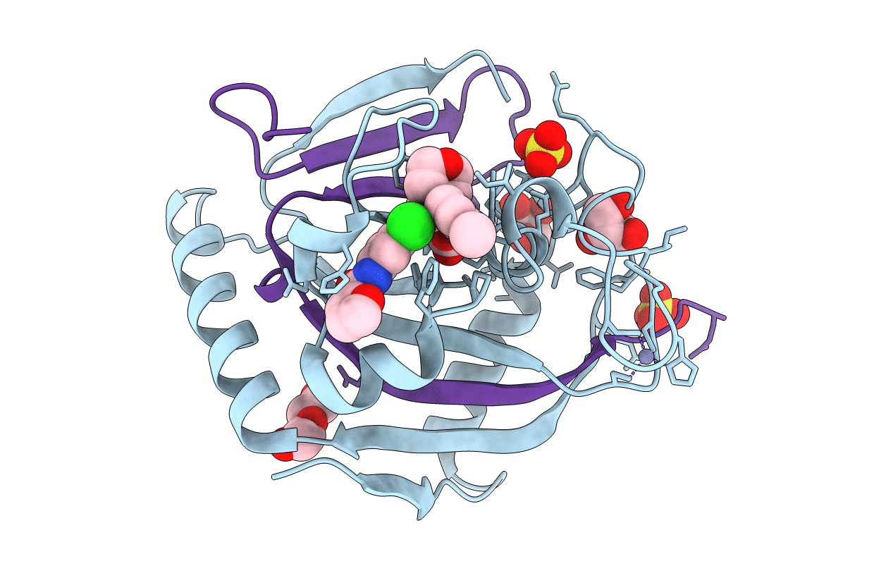
Crystal structure of human tankyrase 2 in complex with OD39
Biological Source:
Source Organism:
PDB Version:
Deposition Date:
2015-08-24
Release Date:
2016-01-13
Method Details:
Experimental Method:
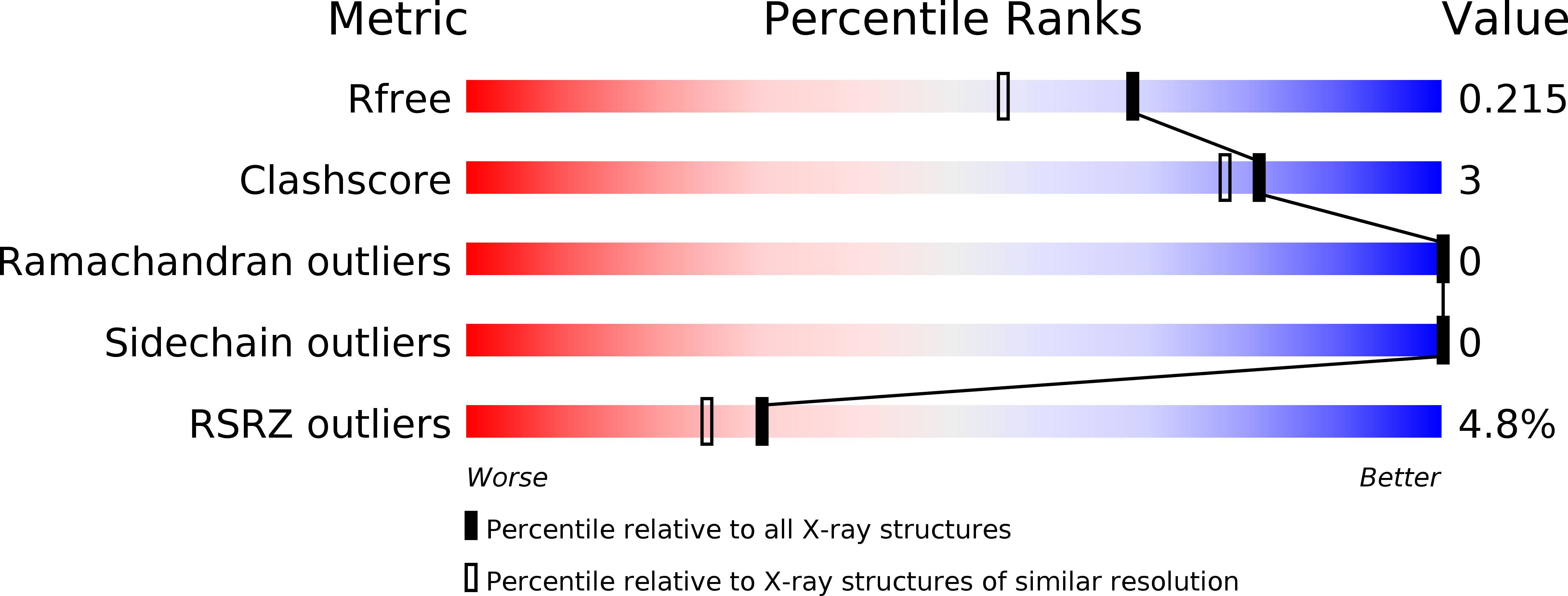
Resolution:
1.80 Å
R-Value Free:
0.20
R-Value Work:
0.16
R-Value Observed:
0.16
Space Group:
P 41 21 2
Macromolecular Entities
Polymer Type:polypeptide(L)
Description:TANKYRASE-2
Chain IDs:A
Chain Length:191
Number of Molecules:1
Biological Source:HOMO SAPIENS
Polymer Type:polypeptide(L)
Description:TANKYRASE-2
Chain IDs:B
Chain Length:48
Number of Molecules:1
Biological Source:HOMO SAPIENS
Primary Citation
Development and Structural Analysis of Adenosine Site Binding Tankyrase Inhibitors.
Bioorg.Med.Chem.Lett. 26 328 ? (2016)
PMID: 26706174 DOI: 10.1016/J.BMCL.2015.12.018

Abstact

Tankyrases 1 and 2, the specialized members of the ARTD protein family, are druggable biotargets whose inhibition may have therapeutic potential against cancer, metabolic disease, fibrotic disease, fibrotic wound healing and HSV viral infections. We have previously identified a novel tankyrase inhibitor scaffold, JW55, and showed that it reduces mouse colon adenoma formation in vivo. Here we expanded the scaffold and profiled the selectivity of the compounds against a panel of human ARTDs. The scaffold also enables a fine modulation of selectivity towards either tankyrase 1 or tankyrase 2. In order to get insight about the binding mode of the inhibitors, we solved crystal structures of the compounds in complex with tankyrase 2. The compounds bind to the adenosine pocket of the catalytic domain and cause changes in the protein structure that are modulated by the chemical modifications of the compounds. The structural analysis allows further rational development of this compound class as a potent and selective tankyrase inhibitor.

Legend

Protein

Chemical

Disease

Primary Citation of related structures