Deposition Date
2015-07-21
Release Date
2015-09-09
Last Version Date
2024-11-13
Entry Detail
PDB ID:
5A9I
Keywords:
Title:
Crystal structure of the extracellular domain of PepT2
Biological Source:
Source Organism(s):
RATTUS NORVEGICUS (Taxon ID: 10116)
Expression System(s):
Method Details:
Experimental Method:
Resolution:
2.84 Å
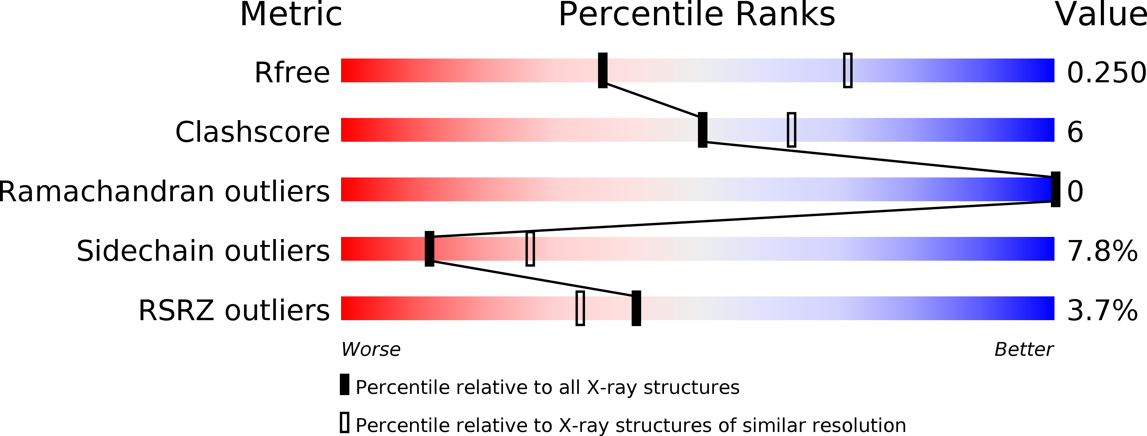
R-Value Free:
0.24
R-Value Work:
0.19
R-Value Observed:
0.19
Space Group:
P 32 2 1