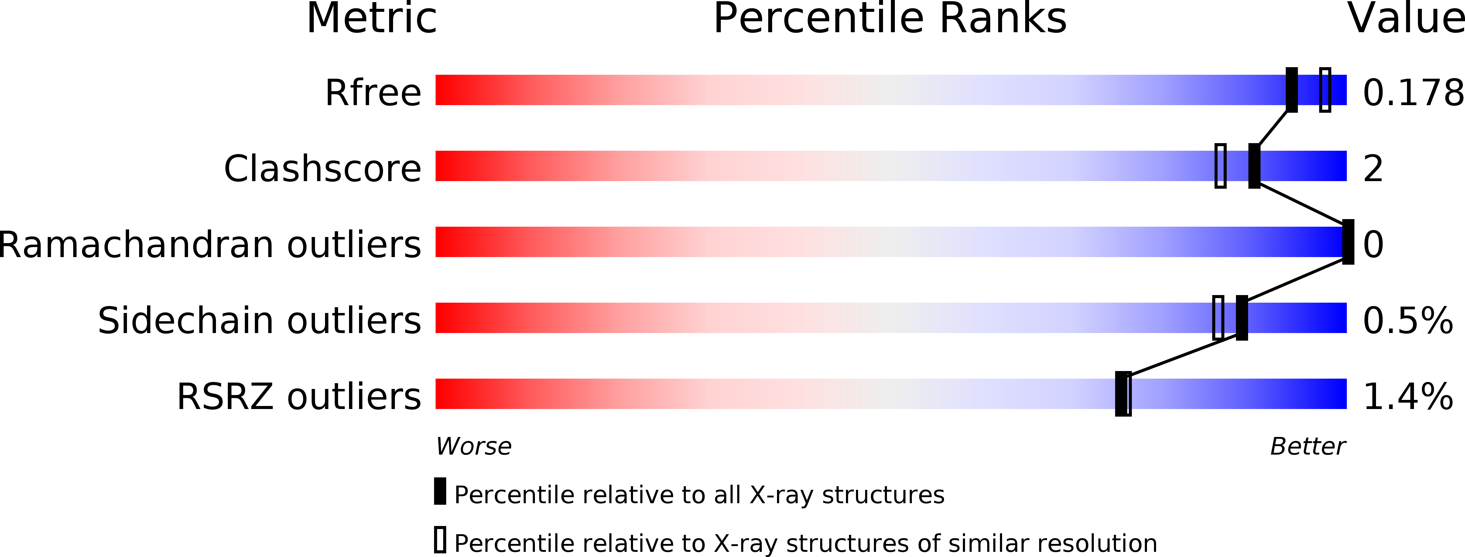
5A8M image
Entry Detail
PDB ID:
5A8M
Keywords:
Title:
Crystal structure of the selenomethionine derivative of beta-glucanase SdGluc5_26A from Saccharophagus degradans
Biological Source:
Host Organism:
PDB Version:
Deposition Date:
2015-07-16
Release Date:
2016-01-20
Method Details:
Experimental Method:
Resolution:
1.86 Å
R-Value Free:
0.16
R-Value Work:
0.14
R-Value Observed:
0.14
Space Group:
P 41 21 2
Macromolecular Entities
Polymer Type:polypeptide(L)
Description:PUTATIVE RETAINING B-GLYCOSIDASE
Mutations:YES
Chain IDs:A, B, C
Chain Length:365
Number of Molecules:3
Biological Source:SACCHAROPHAGUS DEGRADANS 2-40
Modified Residue
Compound ID Chain ID Parent Comp ID Details 2D Image
MSE A MET SELENOMETHIONINE
Primary Citation
The Quaternary Structure of a Glycoside Hydrolase Dictates Specificity Towards Beta-Glucans
J.Biol.Chem. 291 7183 ? (2016)
PMID: 26755730 DOI: 10.1074/JBC.M115.695999

Abstact

In the Carbohydrate-Active Enzyme (CAZy) database, glycoside hydrolase family 5 (GH5) is a large family with more than 6,000 sequences. Among the 51 described GH5 subfamilies, subfamily GH5_26 contains members that display either endo-β(1,4)-glucanase or β(1,3;1,4)-glucanase activities. In this study, we focused on the GH5_26 enzyme fromSaccharophagus degradans(SdGluc5_26A), a marine bacterium known for its capacity to degrade a wide diversity of complex polysaccharides.SdGluc5_26A displays lichenase activity toward β(1,3;1,4)-glucans with a side cellobiohydrolase activity toward β(1,4)-glucans. The three-dimensional structure ofSdGluc5_26A adopts a stable trimeric quaternary structure also observable in solution. The N-terminal region ofSdGluc5_26A protrudes into the active site of an adjacent monomer. To understand whether this occupation of the active site could influence its activity, we conducted a comprehensive enzymatic characterization ofSdGluc5_26A and of a mutant truncated at the N terminus. Ligand complex structures and kinetic analyses reveal that the N terminus governs the substrate specificity ofSdGluc5_26A. Its deletion opens the enzyme cleft at the -3 subsite and turns the enzyme into an endo-β(1,4)-glucanase. This study demonstrates that experimental approaches can reveal structure-function relationships out of reach of current bioinformatic predictions.

Legend

Protein

Chemical

Disease

Primary Citation of related structures