5A8B image
Deposition Date 2015-07-14
Release Date 2016-02-10
Last Version Date 2024-01-10
Entry Detail
PDB ID:
5A8B
Title:
Structure of a parallel dimer of the aureochrome 1a LOV domain from Phaeodactylum tricornutum
Biological Source:
Source Organism:
Host Organism:
Method Details:
Experimental Method:
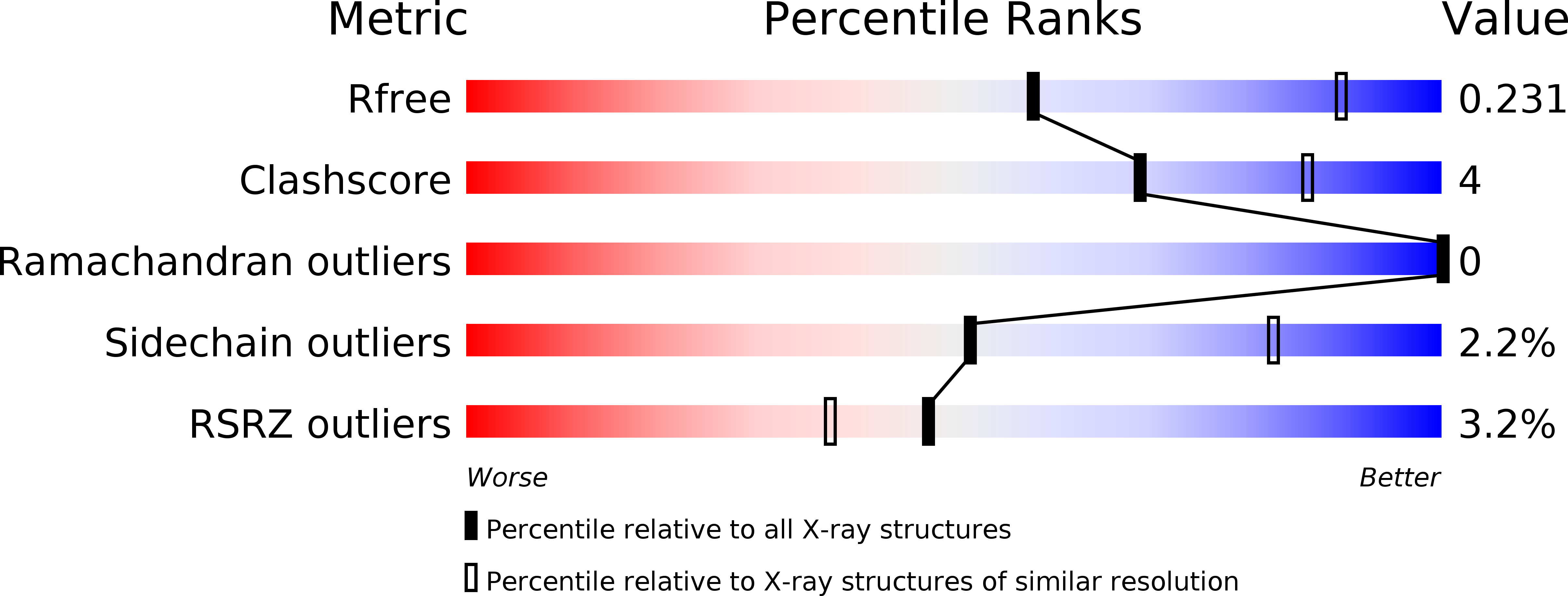
Resolution:
2.79 Å
R-Value Free:
0.23
R-Value Work:
0.17
R-Value Observed:
0.17
Space Group:
P 1 21 1
Macromolecular Entities
Structures with similar UniProt ID
Protein Blast
Polymer Type:polypeptide(L)
Molecule:PTAUREO1A LOV2 DOMAIN
Chain IDs:A, B, C, D
Chain Length:162
Number of Molecules:4
Biological Source:PHAEODACTYLUM TRICORNUTUM
Primary Citation
Structure of a Native-Like Aureochrome 1A Lov Domain Dimer from Phaeodactylum Tricornutum.
Structure 24 171 ? (2016)
PMID: 26688213 DOI: 10.1016/J.STR.2015.10.022

Abstact

Light-oxygen-voltage (LOV) domains absorb blue light for mediating various biological responses in all three domains of life. Aureochromes from stramenopile algae represent a subfamily of photoreceptors that differs by its inversed topology with a C-terminal LOV sensor and an N-terminal effector (basic region leucine zipper, bZIP) domain. We crystallized the LOV domain including its flanking helices, A'α and Jα, of aureochrome 1a from Phaeodactylum tricornutum in the dark state and solved the structure at 2.8 Å resolution. Both flanking helices contribute to the interface of the native-like dimer. Small-angle X-ray scattering shows light-induced conformational changes limited to the dimeric envelope as well as increased flexibility in the lit state for the flanking helices. These rearrangements are considered to be crucial for the formation of the light-activated dimer. Finally, the LOV domain of the class 2 aureochrome PtAUREO2 was shown to lack a chromophore because of steric hindrance caused by M301.

Legend

Protein

Chemical

Disease

Primary Citation of related structures
Feedback Form
Name
Email
Institute
Feedback