
Deposition Date
2015-07-02
Release Date
2016-05-25
Last Version Date
2024-01-10
Entry Detail
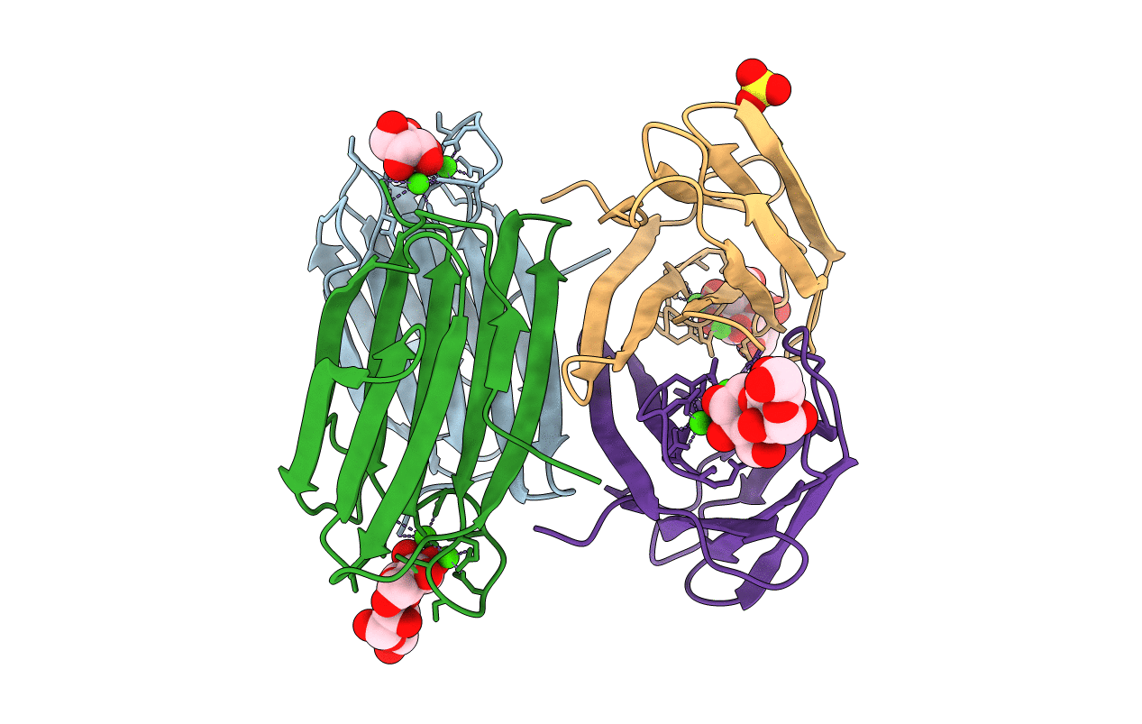
PDB ID:
5A6Y
Keywords:
Title:
Structure of the LecB lectin from Pseudomonas aeruginosa strain PA14 in complex with mannose-alpha1,3mannoside
Biological Source:
Source Organism(s):
PSEUDOMONAS AERUGINOSA (Taxon ID: 208963)
Expression System(s):
Method Details:
Experimental Method:
Resolution:
1.40 Å
R-Value Free:
0.16
R-Value Work:
0.12
R-Value Observed:
0.12
Space Group:
P 1 21 1
