5A4G image
Entry Detail
PDB ID:
5A4G
Title:
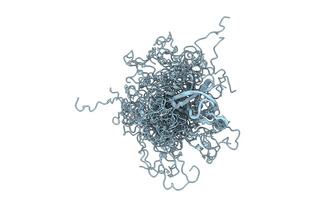
NMR structure of a 180 residue construct encompassing the N-terminal metal-binding site and the membrane proximal domain of SilB from Cupriavidus metallidurans CH34
Biological Source:
Source Organism:
Host Organism:
PDB Version:
Deposition Date:
2015-06-09
Release Date:
2016-05-18
Method Details:
Experimental Method:
Conformers Calculated:
1000
Conformers Submitted:
20
Selection Criteria:
TOTAL ENERGY
Macromolecular Entities
Polymer Type:polypeptide(L)
Description:SILB, SILVER EFFLUX PROTEIN, MFP COMPONENT OF THE THREE COMPONENTS PROTON ANTIPORTER METAL EFFLUX SYSTEM
Chain IDs:A
Chain Length:180
Number of Molecules:1
Biological Source:CUPRIAVIDUS METALLIDURANS
Ligand Molecules
Primary Citation
Structural and Functional Investigation of the Ag+/Cu+-Binding Domains of the Periplasmic Adaptor Protein Silb from Cupriavidus Metallidurans Ch34.
Biochemistry 55 2883 ? (2016)
PMID: 27145046 DOI: 10.1021/ACS.BIOCHEM.6B00022

Abstact

Silver ion resistance in bacteria mainly relies on efflux systems, and notably on tripartite efflux complexes involving a transporter from the resistance-nodulation-cell division (RND) superfamily, such as the SilCBA system from Cupriavidus metallidurans CH34. The periplasmic adaptor protein SilB hosts two specific metal coordination sites, located in the N-terminal and C-terminal domains, respectively, that are believed to play a different role in the efflux mechanism and the trafficking of metal ions from the periplasm to the RND transporter. On the basis of the known domain structure of periplasmic adaptor proteins, we designed different protein constructs derived from SilB domains with either one or two metal binding sites per protein chain. ITC data acquired on proteins with single metal sites suggest a slightly higher affinity of Ag(+) for the N-terminal metal site, compared to that for the C-terminal one. Remarkably, via the study of a protein construct featuring both metal sites, nuclear magnetic resonance (NMR) and fluorescence spectroscopies concordantly show that the C-terminal site is saturated prior to the N-terminal one. The C-terminal binding site is supposed to transfer the metal ions to the RND protein, while the transport driven by this latter is activated upon binding of the metal ion to the N-terminal site. Our results suggest that the filling of the C-terminal metal site is a key prerequisite for preventing futile activation of the transport system. Exhaustive NMR studies reveal for the first time the structure and dynamics of the functionally important N-terminal domain connected to the membrane proximal domain as well as of its Ag(+) binding site.

Legend

Protein

Chemical

Disease

Primary Citation of related structures