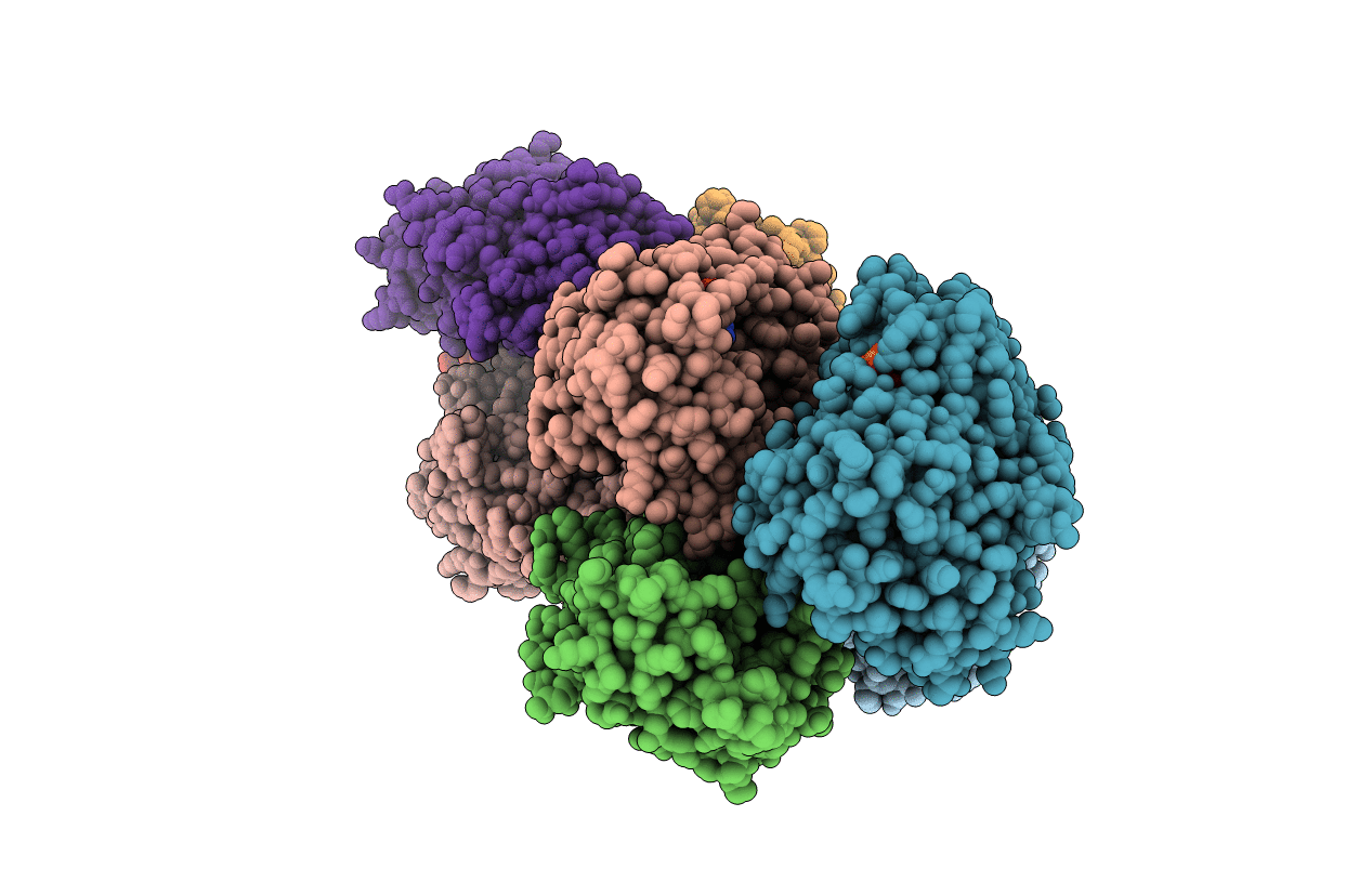
5A4D image
Entry Detail
PDB ID:
5A4D
Keywords:
Title:
Crystal structure of the chloroplastic gamma-ketol reductase from Arabidopsis thaliana bound to 13KOTE and NADP
Biological Source:
Source Organism:
Host Organism:
PDB Version:
Deposition Date:
2015-06-08
Release Date:
2016-09-28
Method Details:
Experimental Method:
Resolution:
2.81 Å
R-Value Free:
0.22
R-Value Work:
0.18
R-Value Observed:
0.19
Space Group:
P 1 21 1
Macromolecular Entities
Polymer Type:polypeptide(L)
Description:PUTATIVE QUINONE-OXIDOREDUCTASE HOMOLOG, CHLOROPLASTIC
Chain IDs:A, B, C, D, E, F, G, H
Chain Length:329
Number of Molecules:8
Biological Source:ARABIDOPSIS THALIANA
Primary Citation
Crystal Structure of the Chloroplastic Oxoene Reductase ceQORH from Arabidopsis thaliana.
Front Plant Sci 8 329 329 (2017)
PMID: 28337214 DOI: 10.3389/fpls.2017.00329

Abstact

Enzymatic and non-enzymatic peroxidation of polyunsaturated fatty acids give rise to accumulation of aldehydes, ketones, and α,β-unsaturated carbonyls of various lengths, known as oxylipins. Oxylipins with α,β-unsaturated carbonyls are reactive electrophile species and are toxic. Cells have evolved several mechanisms to scavenge reactive electrophile oxylipins and decrease their reactivity such as by coupling with glutathione, or by reduction using NAD(P)H-dependent reductases and dehydrogenases of various substrate specificities. Plant cell chloroplasts produce reactive electrophile oxylipins named γ-ketols downstream of enzymatic lipid peroxidation. The chloroplast envelope quinone oxidoreductase homolog (ceQORH) from Arabidopsis thaliana was previously shown to reduce the reactive double bond of γ-ketols. In marked difference with its cytosolic homolog alkenal reductase (AtAER) that displays a high activity toward the ketodiene 13-oxo-9(Z),11(E)-octadecadienoic acid (13-KODE) and the ketotriene 13-oxo-9(Z), 11(E), 15(Z)-octadecatrienoic acid (13-KOTE), ceQORH binds, but does not reduce, 13-KODE and 13-KOTE. Crystal structures of apo-ceQORH and ceQORH bound to 13-KOTE or to NADP+ and 13-KOTE have been solved showing a large ligand binding site, also observed in the structure of the cytosolic alkenal/one reductase. Positioning of the α,β-unsaturated carbonyl of 13-KOTE in ceQORH-NADP+-13-KOTE, far away from the NADP+ nicotinamide ring, provides a rational for the absence of activity with the ketodienes and ketotrienes. ceQORH is a monomeric enzyme in solution whereas other enzymes from the quinone oxidoreductase family are stable dimers and a structural explanation of this difference is proposed. A possible in vivo role of ketodienes and ketotrienes binding to ceQORH is also discussed.

Legend

Protein

Chemical

Disease

Primary Citation of related structures