5A3R image
Deposition Date 2015-06-02
Release Date 2016-04-13
Last Version Date 2024-11-06
Entry Detail
PDB ID:
5A3R
Keywords:
Title:
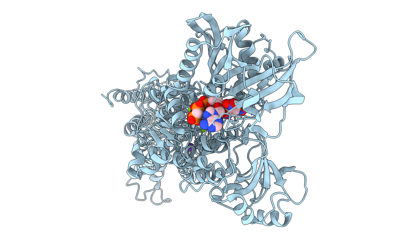
Crystal structure of the (SR) Calcium ATPase E2.BeF3- complex bound to TNP-AMPPCP
Biological Source:
Source Organism:
Method Details:
Experimental Method:
Resolution:
3.05 Å
R-Value Free:
0.25
R-Value Work:
0.21
R-Value Observed:
0.21
Space Group:
P 21 21 21
Macromolecular Entities
Polymer Type:polypeptide(L)
Molecule:SARCOPLASMIC/ENDOPLASMIC RETICULUM CALCIUM ATPASE 1
Gene (Uniprot):ATP2A1
Chain IDs:A
Chain Length:994
Number of Molecules:1
Biological Source:ORYCTOLAGUS CUNICULUS
Modified Residue
Compound ID Chain ID Parent Comp ID Details 2D Image
BFD A ASP ASPARTATE BERYLLIUM TRIFLUORIDE
Primary Citation
Crystal Structure of the Vanadate-Inhibited Ca(2+)-ATPase.
Structure 24 617 ? (2016)
PMID: 27050689 DOI: 10.1016/J.STR.2016.02.018

Abstact

Vanadate is the hallmark inhibitor of the P-type ATPase family; however, structural details of its inhibitory mechanism have remained unresolved. We have determined the crystal structure of sarcoplasmic reticulum Ca(2+)-ATPase with bound vanadate in the absence of Ca(2+). Vanadate is bound at the catalytic site as a planar VO3(-) in complex with water and Mg(2+) in a dephosphorylation transition-state-like conformation. Validating bound VO3(-) by anomalous difference Fourier maps using long-wavelength data we also identify a hitherto undescribed Cl(-) site near the dephosphorylation site. Crystallization was facilitated by trinitrophenyl (TNP)-derivatized nucleotides that bind with the TNP moiety occupying the binding pocket that normally accommodates the adenine of ATP, rationalizing their remarkably high affinity for E2P-like conformations of the Ca(2+)-ATPase. A comparison of the configurations of bound nucleotide analogs in the E2·VO3(-) structure with that in E2·BeF3(-) (E2P ground state analog) reveals multiple binding modes to the Ca(2+)-ATPase.

Legend

Protein

Chemical

Disease

Primary Citation of related structures