5A1J image
Deposition Date 2015-04-30
Release Date 2015-05-13
Last Version Date 2024-01-10
Entry Detail
PDB ID:
5A1J
Title:
Periplasmic Binding Protein CeuE in complex with ferric 4-LICAM
Biological Source:
Source Organism:
Host Organism:
Method Details:
Experimental Method:
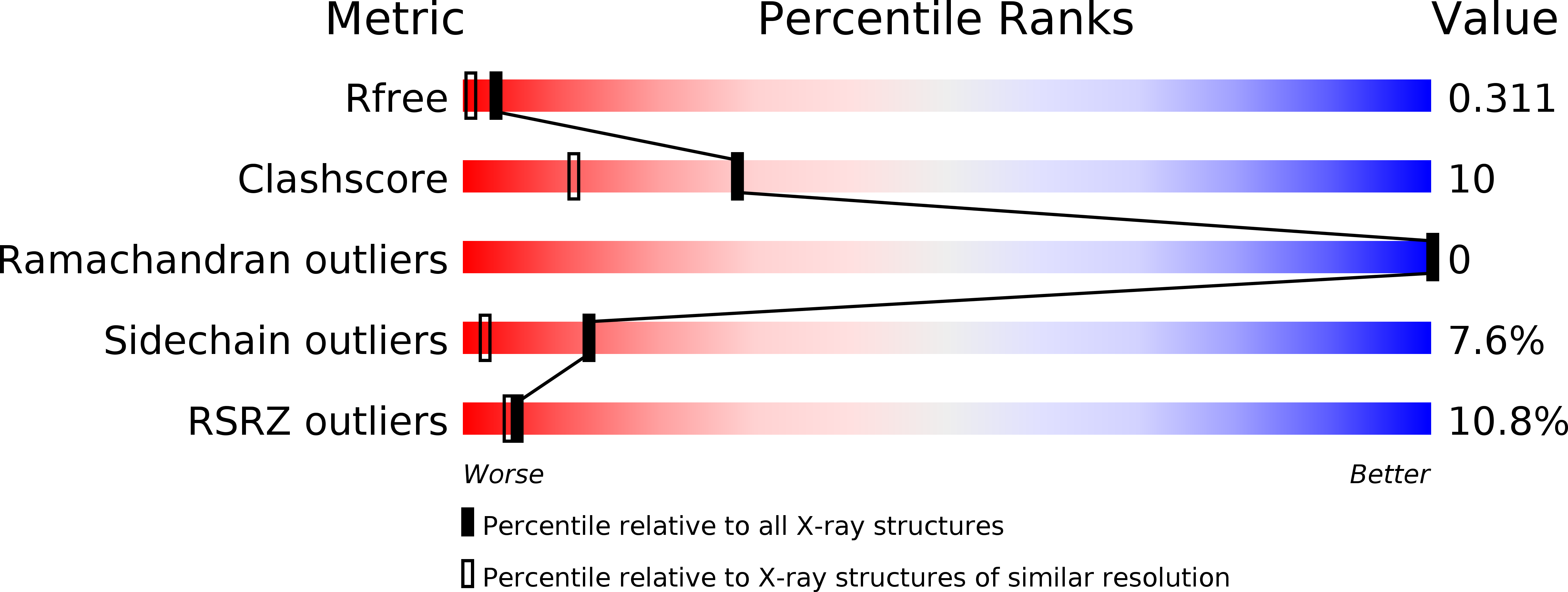
Resolution:
1.60 Å
R-Value Free:
0.31
R-Value Work:
0.26
R-Value Observed:
0.26
Space Group:
P 21 21 21
Macromolecular Entities
Polymer Type:polypeptide(L)
Molecule:ENTEROCHELIN UPTAKE PERIPLASMIC BINDING PROTEIN
Gene (Uniprot):ceuE
Chain IDs:A
Chain Length:288
Number of Molecules:1
Biological Source:CAMPYLOBACTER JEJUNI
Primary Citation
Interactions of a Periplasmic Binding Protein with a Tetradentate Siderophore Mimic.
Angew.Chem.Int.Ed.Engl. 52 4595 ? (2013)
PMID: 23512642 DOI: 10.1002/ANIE.201300751

Abstact

Iron-bound structure: The ferric complex of a tetradentate siderophore mimic was synthesized and co-crystallized with the periplasmic binding protein CeuE of Campylobacter jejuni. In addition to electrostatic and hydrogen-bonding interactions between the binding pocket and the substrate, the structure showed direct coordination of two amino acid side chains to the Fe(III) center (orange, see figure).

Legend

Protein

Chemical

Disease

Primary Citation of related structures