5A16 image
Entry Detail
PDB ID:
5A16
Keywords:
Title:
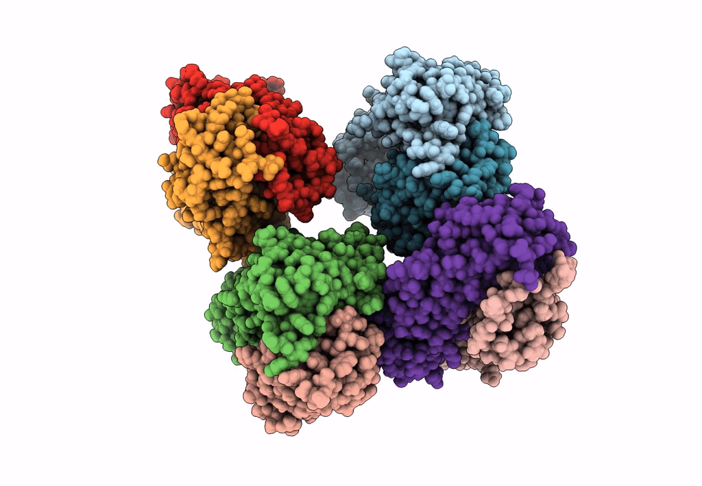
Crystal structure of Fab4201 raised against Human Erythrocyte Anion Exchanger 1
Biological Source:
Source Organism:
PDB Version:
Deposition Date:
2015-04-28
Release Date:
2015-06-10
Method Details:
Experimental Method:
Resolution:
2.50 Å
R-Value Free:
0.25
R-Value Work:
0.21
R-Value Observed:
0.21
Space Group:
P 41
Macromolecular Entities
Polymer Type:polypeptide(L)
Description:FAB4201 HEAVY CHAIN
Chain IDs:A, C, E, G
Chain Length:236
Number of Molecules:4
Biological Source:MUS MUSCULUS
Polymer Type:polypeptide(L)
Description:FAB4201 HEAVY CHAIN
Chain IDs:B, D, F, H
Chain Length:218
Number of Molecules:4
Biological Source:MUS MUSCULUS
Primary Citation
Crystal Structure of the Anion Exchanger Domain of Human Erythrocyte Band 3
Science 350 680 ? (2015)
PMID: 26542571 DOI: 10.1126/SCIENCE.AAA4335

Abstact

Anion exchanger 1 (AE1), also known as band 3 or SLC4A1, plays a key role in the removal of carbon dioxide from tissues by facilitating the exchange of chloride and bicarbonate across the plasma membrane of erythrocytes. An isoform of AE1 is also present in the kidney. Specific mutations in human AE1 cause several types of hereditary hemolytic anemias and/or distal renal tubular acidosis. Here we report the crystal structure of the band 3 anion exchanger domain (AE1(CTD)) at 3.5 angstroms. The structure is locked in an outward-facing open conformation by an inhibitor. Comparing this structure with a substrate-bound structure of the uracil transporter UraA in an inward-facing conformation allowed us to identify the anion-binding position in the AE1(CTD), and to propose a possible transport mechanism that could explain why selected mutations lead to disease.

Legend

Protein

Chemical

Disease

Primary Citation of related structures