4r8v image
Deposition Date 2014-09-03
Release Date 2015-04-15
Last Version Date 2023-11-08
Entry Detail
PDB ID:
4R8V
Keywords:
Title:
Crystal structure of the hydrolase domain of 10-formyltetrahydrofolate dehydrogenase (wild-type) complex with formate
Biological Source:
Source Organism:
Danio rerio (Taxon ID: 7955)
Host Organism:
Method Details:
Experimental Method:
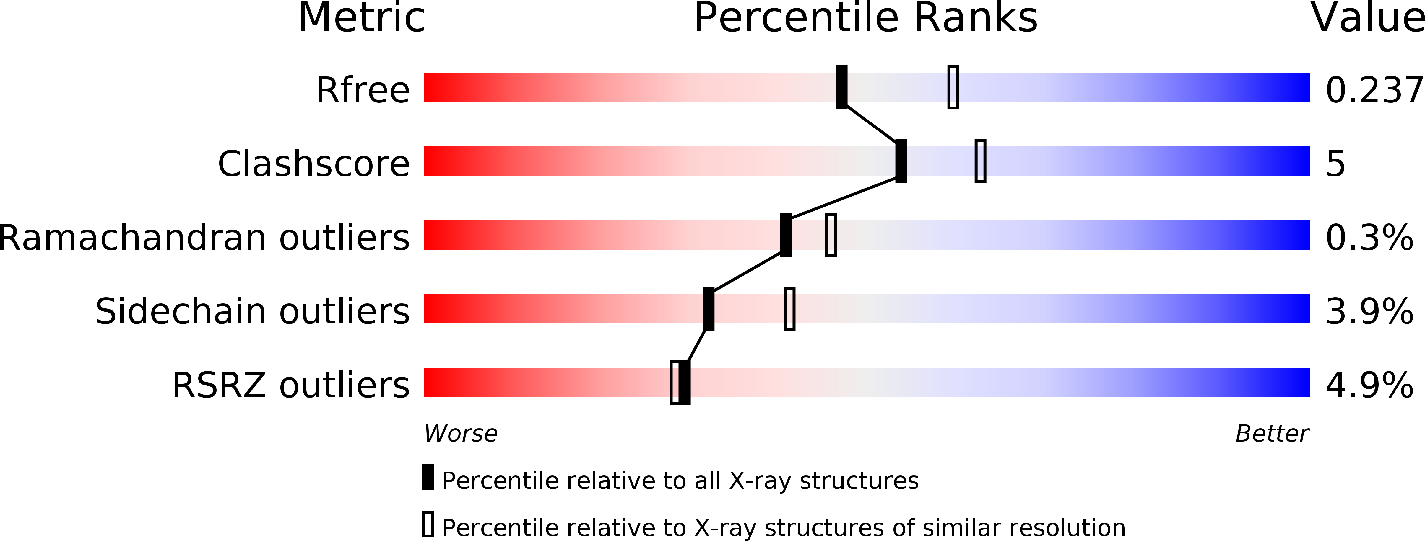
Resolution:
2.20 Å
R-Value Free:
0.23
R-Value Work:
0.19
R-Value Observed:
0.19
Space Group:
P 21 21 2
Macromolecular Entities
Structures with similar UniProt ID
Protein Blast
Polymer Type:polypeptide(L)
Molecule:10-formyltetrahydrofolate dehydrogenase
Gene (Uniprot):aldh1l1
Chain IDs:A
Chain Length:318
Number of Molecules:1
Biological Source:Danio rerio
Primary Citation
Structures of the hydrolase domain of zebrafish 10-formyltetrahydrofolate dehydrogenase and its complexes reveal a complete set of key residues for hydrolysis and product inhibition.
Acta Crystallogr.,Sect.D 71 1006 1021 (2015)
PMID: 25849409 DOI: 10.1107/S1399004715002928

Abstact

10-Formyltetrahydrofolate dehydrogenase (FDH), which is composed of a small N-terminal domain (Nt-FDH) and a large C-terminal domain, is an abundant folate enzyme in the liver and converts 10-formyltetrahydrofolate (10-FTHF) to tetrahydrofolate (THF) and CO2. Nt-FDH alone possesses a hydrolase activity, which converts 10-FTHF to THF and formate in the presence of β-mercaptoethanol. To elucidate the catalytic mechanism of Nt-FDH, crystal structures of apo-form zNt-FDH from zebrafish and its complexes with the substrate analogue 10-formyl-5,8-dideazafolate (10-FDDF) and with the products THF and formate have been determined. The structures reveal that the conformations of three loops (residues 86-90, 135-143 and 200-203) are altered upon ligand (10-FDDF or THF) binding in the active site. The orientations and geometries of key residues, including Phe89, His106, Arg114, Asp142 and Tyr200, are adjusted for substrate binding and product release during catalysis. Among them, Tyr200 is especially crucial for product release. An additional potential THF binding site is identified in the cavity between two zNt-FDH molecules, which might contribute to the properties of product inhibition and THF storage reported for FDH. Together with mutagenesis studies and activity assays, the structures of zNt-FDH and its complexes provide a coherent picture of the active site and a potential THF binding site of zNt-FDH along with the substrate and product specificity, lending new insights into the molecular mechanism underlying the enzymatic properties of Nt-FDH.

Legend

Protein

Chemical

Disease

Primary Citation of related structures