4ZYN image
Deposition Date 2015-05-21
Release Date 2015-08-19
Last Version Date 2024-11-13
Entry Detail
PDB ID:
4ZYN
Keywords:
Title:
Crystal Structure of Parkin E3 ubiquitin ligase (linker deletion; delta 86-130)
Biological Source:
Source Organism:
Host Organism:
Method Details:
Experimental Method:
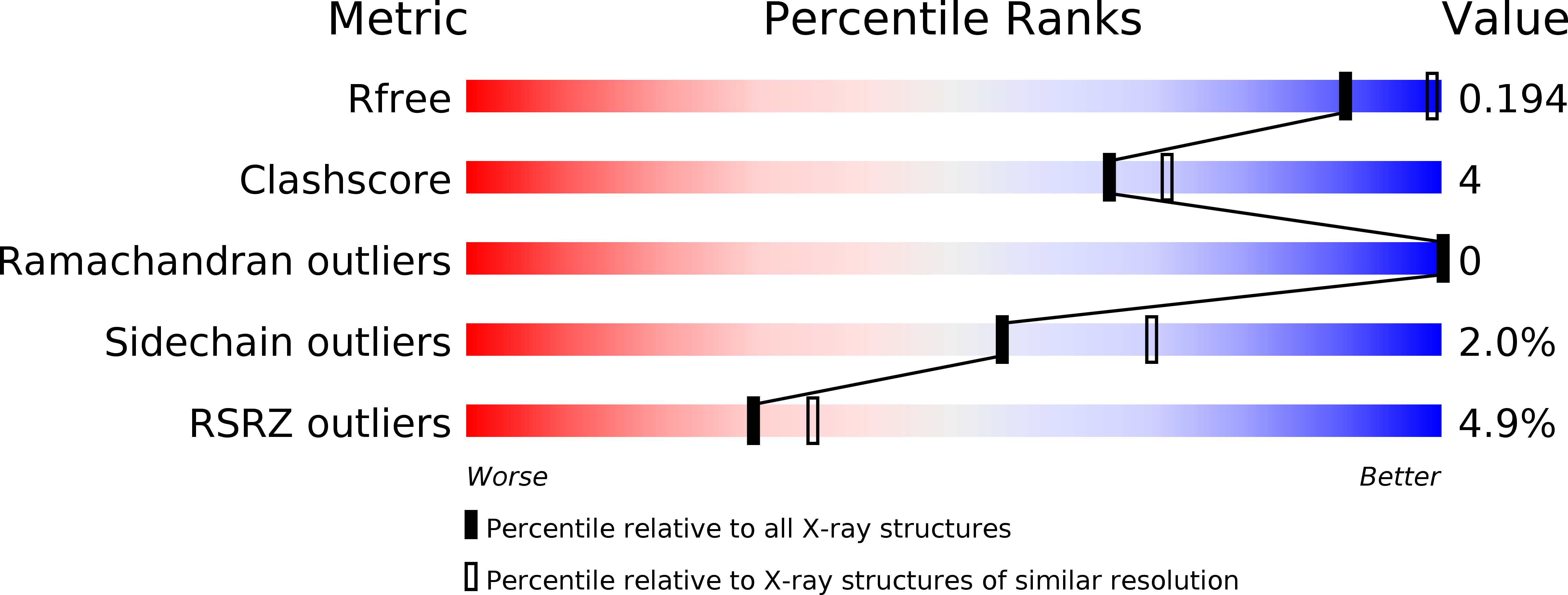
Resolution:
2.54 Å
R-Value Free:
0.23
R-Value Work:
0.19
R-Value Observed:
0.19
Space Group:
H 3
Macromolecular Entities
Polymer Type:polypeptide(L)
Molecule:E3 ubiquitin-protein ligase parkin
Gene (Uniprot):Prkn
Chain IDs:A, B
Chain Length:425
Number of Molecules:2
Biological Source:Rattus norvegicus
Primary Citation
A Ubl/ubiquitin switch in the activation of Parkin.
Embo J. 34 2492 2505 (2015)
PMID: 26254305 DOI: 10.15252/embj.201592237

Abstact

Mutations in Parkin and PINK1 cause an inherited early-onset form of Parkinson's disease. The two proteins function together in a mitochondrial quality control pathway whereby PINK1 accumulates on damaged mitochondria and activates Parkin to induce mitophagy. How PINK1 kinase activity releases the auto-inhibited ubiquitin ligase activity of Parkin remains unclear. Here, we identify a binding switch between phospho-ubiquitin (pUb) and the ubiquitin-like domain (Ubl) of Parkin as a key element. By mutagenesis and SAXS, we show that pUb binds to RING1 of Parkin at a site formed by His302 and Arg305. pUb binding promotes disengagement of the Ubl from RING1 and subsequent Parkin phosphorylation. A crystal structure of Parkin Δ86-130 at 2.54 Å resolution allowed the design of mutations that specifically release the Ubl domain from RING1. These mutations mimic pUb binding and promote Parkin phosphorylation. Measurements of the E2 ubiquitin-conjugating enzyme UbcH7 binding to Parkin and Parkin E3 ligase activity suggest that Parkin phosphorylation regulates E3 ligase activity downstream of pUb binding.

Legend

Protein

Chemical

Disease

Primary Citation of related structures