4ZWJ image
Entry Detail
PDB ID:
4ZWJ
Title:
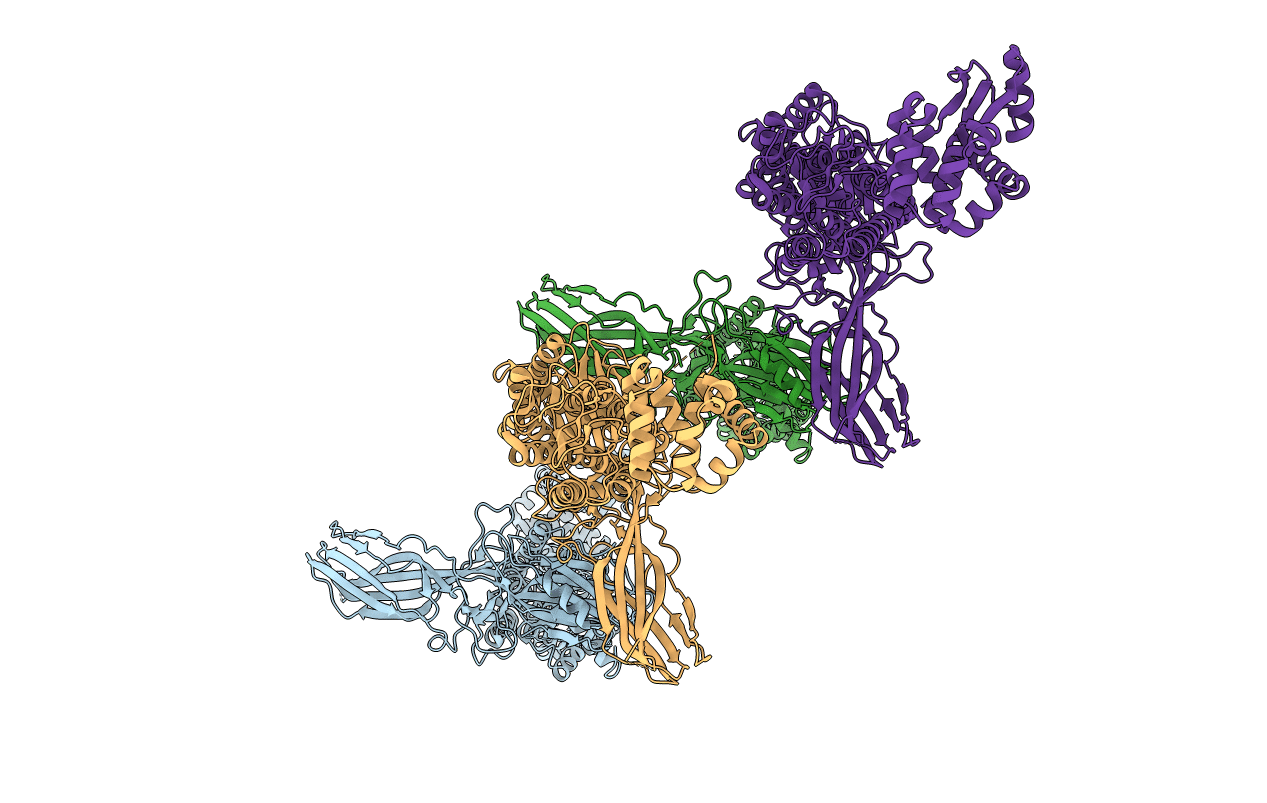
Crystal structure of rhodopsin bound to arrestin by femtosecond X-ray laser
Biological Source:
PDB Version:
Deposition Date:
2015-05-19
Release Date:
2015-07-29
Method Details:
Experimental Method:
Resolution:
3.30 Å
R-Value Free:
0.29
R-Value Work:
0.25
R-Value Observed:
0.25
Space Group:
P 21 21 21
Macromolecular Entities
Polymer Type:polypeptide(L)
Description:Chimera protein of human Rhodopsin, mouse S-arrestin, and T4 Endolysin
Mutations:R12G, C54T, C97A , I137R,N2C, E113Q, M257Y, N282C,L374A, V375A, F376A
Chain IDs:A, B, C, D
Chain Length:906
Number of Molecules:4
Biological Source:Enterobacteria phage T4, Homo sapiens, Mus musculus
Ligand Molecules
Primary Citation

Abstact

G-protein-coupled receptors (GPCRs) signal primarily through G proteins or arrestins. Arrestin binding to GPCRs blocks G protein interaction and redirects signalling to numerous G-protein-independent pathways. Here we report the crystal structure of a constitutively active form of human rhodopsin bound to a pre-activated form of the mouse visual arrestin, determined by serial femtosecond X-ray laser crystallography. Together with extensive biochemical and mutagenesis data, the structure reveals an overall architecture of the rhodopsin-arrestin assembly in which rhodopsin uses distinct structural elements, including transmembrane helix 7 and helix 8, to recruit arrestin. Correspondingly, arrestin adopts the pre-activated conformation, with a ∼20° rotation between the amino and carboxy domains, which opens up a cleft in arrestin to accommodate a short helix formed by the second intracellular loop of rhodopsin. This structure provides a basis for understanding GPCR-mediated arrestin-biased signalling and demonstrates the power of X-ray lasers for advancing the frontiers of structural biology.

Legend

Protein

Chemical

Disease

Primary Citation of related structures