4ZV5 image
Deposition Date 2015-05-18
Release Date 2016-01-27
Last Version Date 2024-11-13
Entry Detail
PDB ID:
4ZV5
Keywords:
Title:
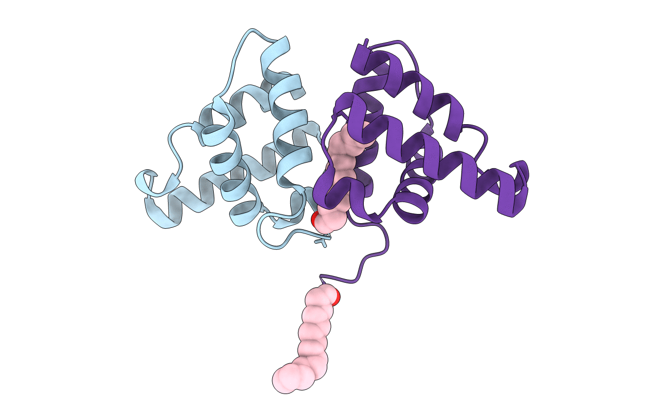
Crystal structure of N-myristoylated mouse mammary tumor virus matrix protein
Biological Source:
Source Organism:
Host Organism:
Method Details:
Experimental Method:
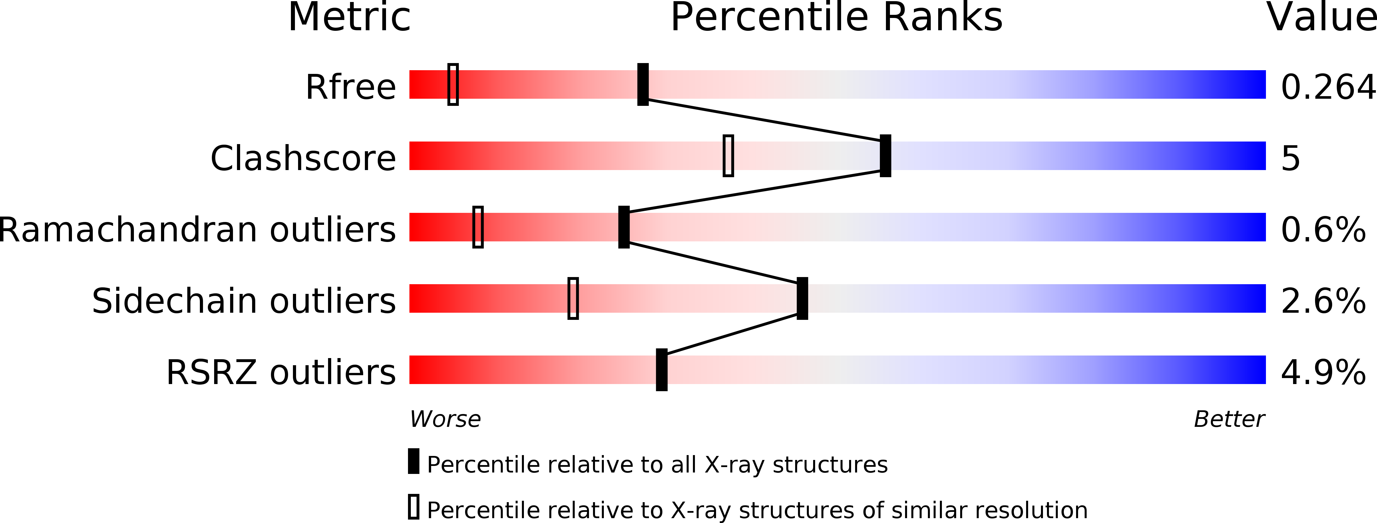
Resolution:
1.57 Å
R-Value Free:
0.26
R-Value Work:
0.22
R-Value Observed:
0.22
Space Group:
P 31 2 1
Macromolecular Entities
Structures with similar UniProt ID
Protein Blast
Polymer Type:polypeptide(L)
Molecule:Matrix protein p10
Chain IDs:A, B
Chain Length:91
Number of Molecules:2
Biological Source:Mouse mammary tumor virus
Ligand Molecules
Primary Citation
Myristoylation drives dimerization of matrix protein from mouse mammary tumor virus.
Retrovirology 13 2 2 (2016)
PMID: 26728401 DOI: 10.1186/s12977-015-0235-8

Abstact

BACKGROUND Myristoylation of the matrix (MA) domain mediates the transport and binding of Gag polyproteins to the plasma membrane (PM) and is required for the assembly of most retroviruses. In betaretroviruses, which assemble immature particles in the cytoplasm, myristoylation is dispensable for assembly but is crucial for particle transport to the PM. Oligomerization of HIV-1 MA stimulates the transition of the myristoyl group from a sequestered to an exposed conformation, which is more accessible for membrane binding. However, for other retroviruses, the effect of MA oligomerization on myristoyl group exposure has not been thoroughly investigated. RESULTS Here, we demonstrate that MA from the betaretrovirus mouse mammary tumor virus (MMTV) forms dimers in solution and that this process is stimulated by its myristoylation. The crystal structure of N-myristoylated MMTV MA, determined at 1.57 Å resolution, revealed that the myristoyl groups are buried in a hydrophobic pocket at the dimer interface and contribute to dimer formation. Interestingly, the myristoyl groups in the dimer are mutually swapped to achieve energetically stable binding, as documented by molecular dynamics modeling. Mutations within the myristoyl binding site resulted in reduced MA dimerization and extracellular particle release. CONCLUSIONS Based on our experimental, structural, and computational data, we propose a model for dimerization of MMTV MA in which myristoyl groups stimulate the interaction between MA molecules. Moreover, dimer-forming MA molecules adopt a sequestered conformation with their myristoyl groups entirely buried within the interaction interface. Although this differs from the current model proposed for lentiviruses, in which oligomerization of MA triggers exposure of myristoyl group, it appears convenient for intracellular assembly, which involves no apparent membrane interaction and allows the myristoyl group to be sequestered during oligomerization.

Legend

Protein

Chemical

Disease

Primary Citation of related structures
Feedback Form
Name
Email
Institute
Feedback