4ZT1 image
Entry Detail
PDB ID:
4ZT1
Keywords:
Title:
Crystal structure of human E-Cadherin (residues 3-213) in x-dimer conformation
Biological Source:
Source Organism:
PDB Version:
Deposition Date:
2015-05-14
Release Date:
2016-06-01
Method Details:
Experimental Method:
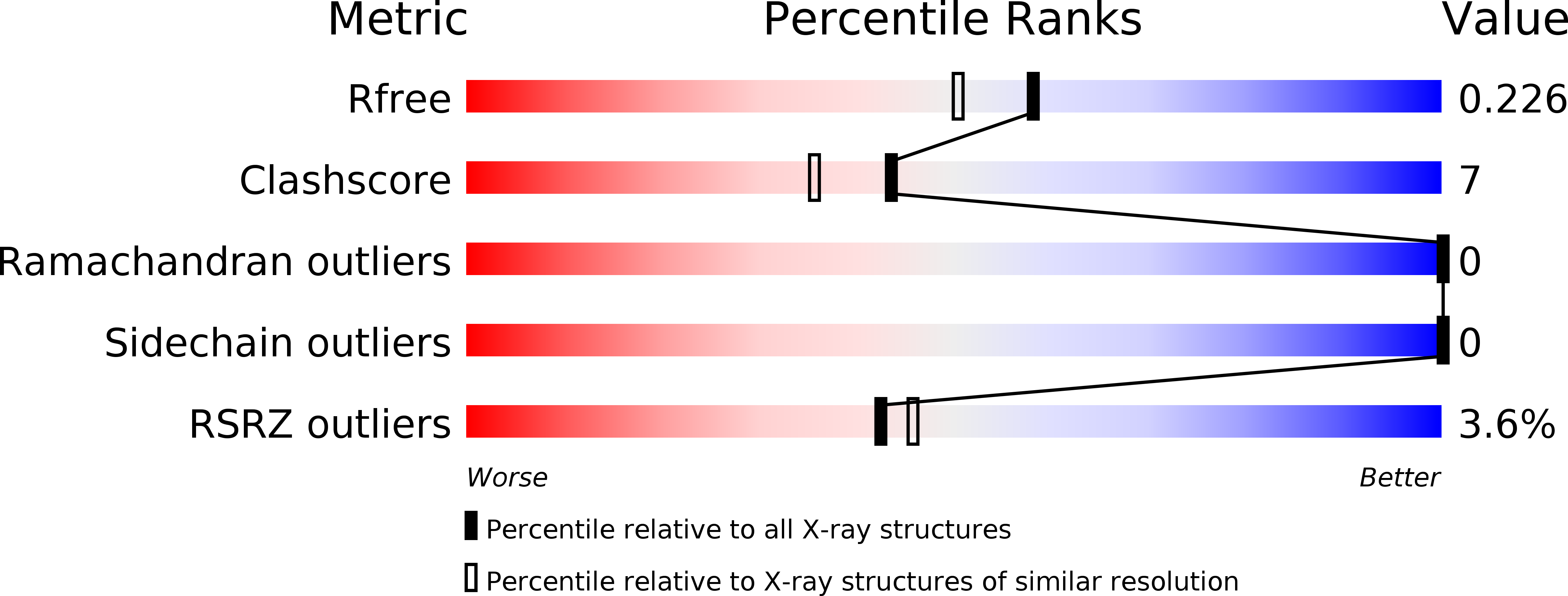
Resolution:
1.92 Å
R-Value Free:
0.21
R-Value Work:
0.19
R-Value Observed:
0.19
Space Group:
C 1 2 1
Macromolecular Entities
Polymer Type:polypeptide(L)
Description:Cadherin-1
Chain IDs:A, B
Chain Length:211
Number of Molecules:2
Biological Source:Homo sapiens
Ligand Molecules
Primary Citation
Crystal Structure of Human E-Cadherin-EC1EC2 in Complex with a Peptidomimetic Competitive Inhibitor of Cadherin Homophilic Interaction.
J.Med.Chem. 59 5089 5094 (2016)
PMID: 27120112 DOI: 10.1021/acs.jmedchem.5b01487

Abstact

Cadherins are transmembrane cell adhesion proteins whose aberrant expression often correlates with cancer development and proliferation. We report the crystal structure of an E-cadherin extracellular fragment in complex with a peptidomimetic compound that was previously shown to partially inhibit cadherin homophilic adhesion. The structure reveals an unexpected binding mode and allows the identification of a druggable cadherin interface, thus paving the way to a future structure-guided design of cell adhesion inhibitors against cadherin-expressing solid tumors.

Legend

Protein

Chemical

Disease

Primary Citation of related structures