4ZRL image
Deposition Date 2015-05-12
Release Date 2015-07-08
Last Version Date 2024-11-20
Entry Detail
PDB ID:
4ZRL
Keywords:
Title:
Structure of the non canonical Poly(A) polymerase complex GLD-2 - GLD-3
Biological Source:
Source Organism:
Host Organism:
Method Details:
Experimental Method:
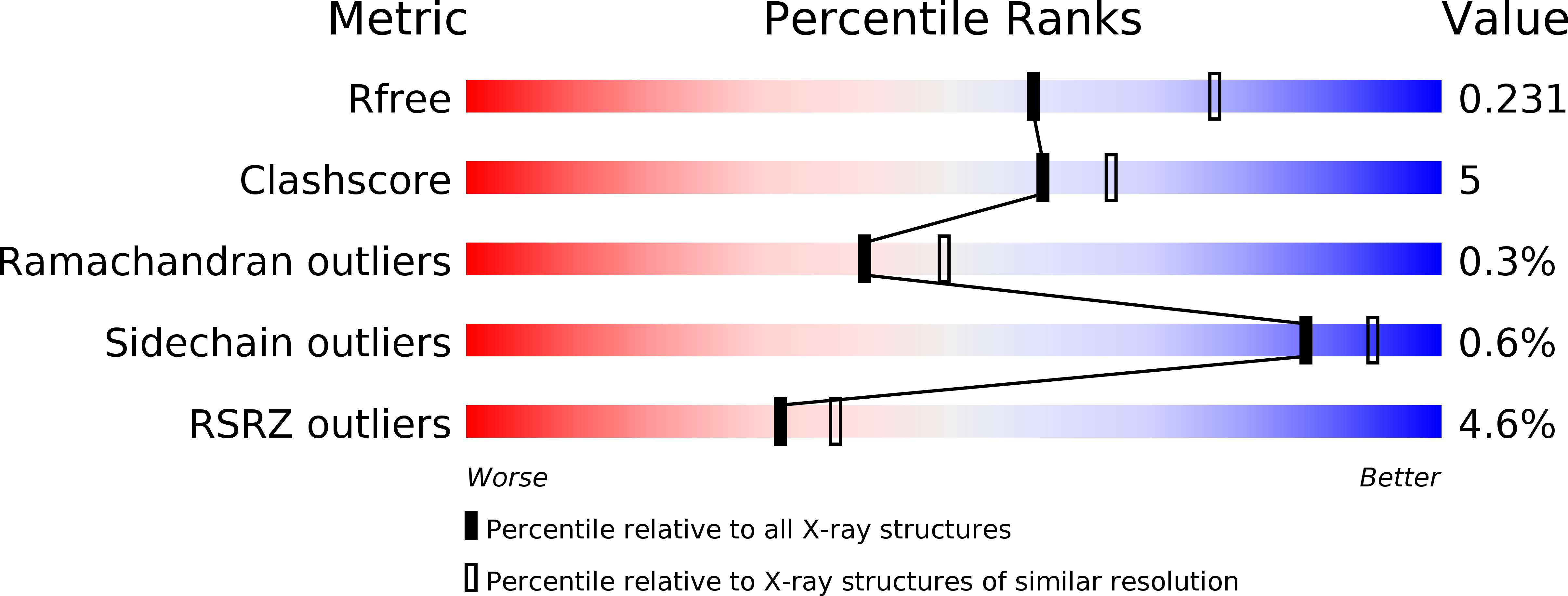
Resolution:
2.28 Å
R-Value Free:
0.23
R-Value Work:
0.18
R-Value Observed:
0.18
Space Group:
P 21 21 21
Macromolecular Entities
Structures with similar UniProt ID
Protein Blast
Polymer Type:polypeptide(L)
Molecule:Poly(A) RNA polymerase gld-2
Gene (Uniprot):gld-2
Chain IDs:A
Chain Length:364
Number of Molecules:1
Biological Source:Caenorhabditis elegans
Structures with similar UniProt ID
Protein Blast
Polymer Type:polypeptide(L)
Molecule:Defective in germ line development protein 3
Gene (Uniprot):gld-3
Chain IDs:B
Chain Length:77
Number of Molecules:1
Biological Source:Caenorhabditis elegans
Modified Residue
Compound ID Chain ID Parent Comp ID Details 2D Image
MSE A MET modified residue
Ligand Molecules
Primary Citation
Structural basis for the activation of the C. elegans noncanonical cytoplasmic poly(A)-polymerase GLD-2 by GLD-3.
Proc.Natl.Acad.Sci.USA 112 8614 8619 (2015)
PMID: 26124149 DOI: 10.1073/pnas.1504648112

Abstact

The Caenorhabditis elegans germ-line development defective (GLD)-2-GLD-3 complex up-regulates the expression of genes required for meiotic progression. GLD-2-GLD-3 acts by extending the short poly(A) tail of germ-line-specific mRNAs, switching them from a dormant state into a translationally active state. GLD-2 is a cytoplasmic noncanonical poly(A) polymerase that lacks the RNA-binding domain typical of the canonical nuclear poly(A)-polymerase Pap1. The activity of C. elegans GLD-2 in vivo and in vitro depends on its association with the multi-K homology (KH) domain-containing protein, GLD-3, a homolog of Bicaudal-C. We have identified a minimal polyadenylation complex that includes the conserved nucleotidyl-transferase core of GLD-2 and the N-terminal domain of GLD-3, and determined its structure at 2.3-Å resolution. The structure shows that the N-terminal domain of GLD-3 does not fold into the predicted KH domain but wraps around the catalytic domain of GLD-2. The picture that emerges from the structural and biochemical data are that GLD-3 activates GLD-2 both indirectly by stabilizing the enzyme and directly by contributing positively charged residues near the RNA-binding cleft. The RNA-binding cleft of GLD-2 has distinct structural features compared with the poly(A)-polymerases Pap1 and Trf4. Consistently, GLD-2 has distinct biochemical properties: It displays unusual specificity in vitro for single-stranded RNAs with at least one adenosine at the 3' end. GLD-2 thus appears to have evolved specialized nucleotidyl-transferase properties that match the 3' end features of dormant cytoplasmic mRNAs.

Legend

Protein

Chemical

Disease

Primary Citation of related structures