4ZQK image
Deposition Date 2015-05-10
Release Date 2015-11-04
Last Version Date 2024-10-16
Entry Detail
PDB ID:
4ZQK
Keywords:
Title:
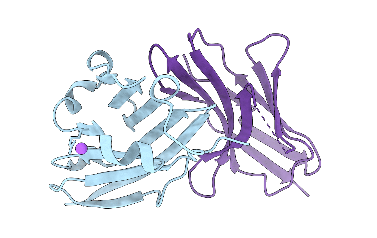
Structure of the complex of human programmed death-1 (PD-1) and its ligand PD-L1.
Biological Source:
Source Organism:
Homo sapiens (Taxon ID: 9606)
Method Details:
Experimental Method:
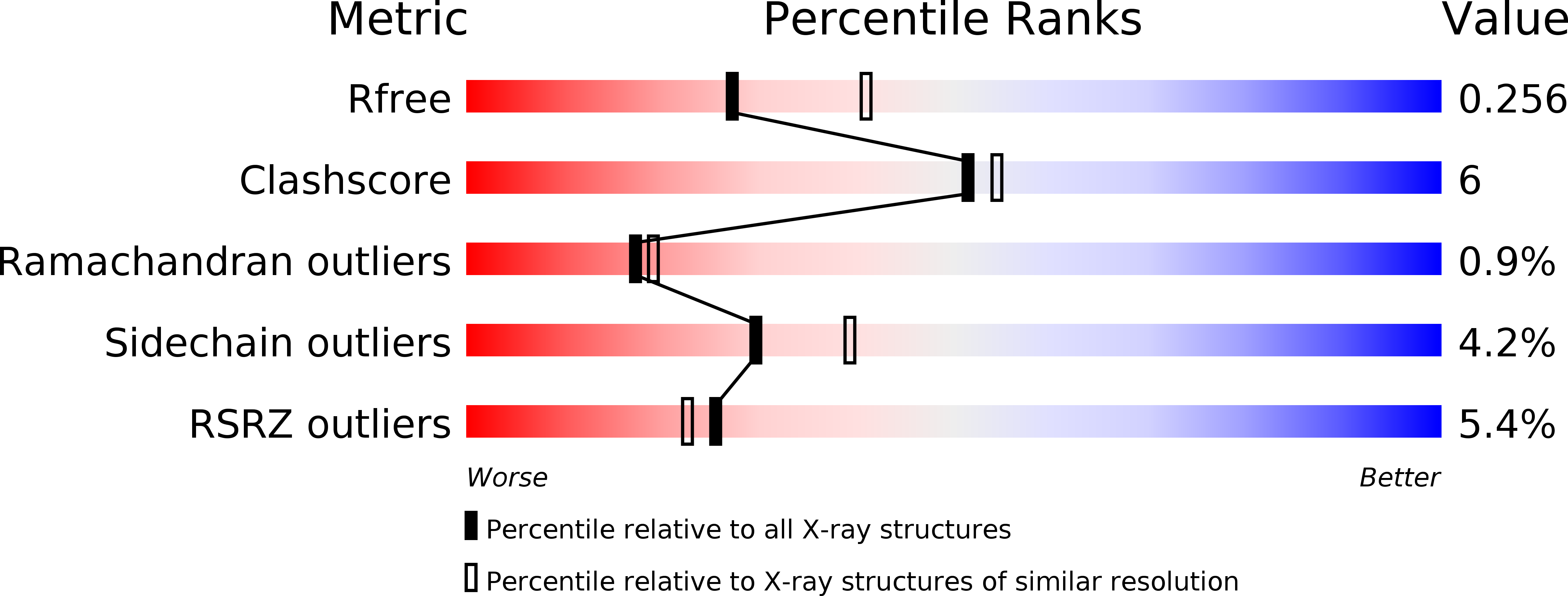
Resolution:
2.45 Å
R-Value Free:
0.25
R-Value Work:
0.20
R-Value Observed:
0.20
Space Group:
P 31 2 1
Macromolecular Entities
Structures with similar UniProt ID
Protein Blast
Polymer Type:polypeptide(L)
Molecule:Programmed cell death 1 ligand 1
Gene (Uniprot):CD274
Chain IDs:A
Chain Length:115
Number of Molecules:1
Biological Source:Homo sapiens
Structures with similar UniProt ID
Protein Blast
Polymer Type:polypeptide(L)
Molecule:Programmed cell death protein 1
Gene (Uniprot):PDCD1
Mutagens:C93S
Chain IDs:B
Chain Length:118
Number of Molecules:1
Biological Source:Homo sapiens
Ligand Molecules
Primary Citation
Structure of the Complex of Human Programmed Death 1, PD-1, and Its Ligand PD-L1.
Structure 23 2341 2348 (2015)
PMID: 26602187 DOI: 10.1016/j.str.2015.09.010

Abstact

Targeting the PD-1/PD-L1 immunologic checkpoint with monoclonal antibodies has recently provided breakthrough progress in the treatment of melanoma, non-small cell lung cancer, and other types of cancer. Small-molecule drugs interfering with this pathway are highly awaited, but their development is hindered by insufficient structural information. This study reveals the molecular details of the human PD-1/PD-L1 interaction based on an X-ray structure of the complex. First, it is shown that the ligand binding to human PD-1 is associated with significant plasticity within the receptor. Second, a detailed molecular map of the interaction surface is provided, allowing definition of the regions within both interacting partners that may likely be targeted by small molecules.

Legend

Protein

Chemical

Disease

Primary Citation of related structures
Feedback Form
Name
Email
Institute
Feedback