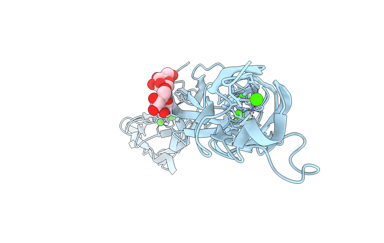
4ZPS image
Deposition Date 2015-05-08
Release Date 2015-10-28
Last Version Date 2024-11-06
Entry Detail
PDB ID:
4ZPS
Keywords:
Title:
Crystal Structure of Protocadherin Gamma A8 EC1-3
Biological Source:
Source Organism:
Mus musculus (Taxon ID: 10090)
Host Organism:
Method Details:
Experimental Method:
Resolution:
2.90 Å
R-Value Free:
0.28
R-Value Work:
0.26
R-Value Observed:
0.26
Space Group:
I 21 21 21
Macromolecular Entities
Polymer Type:polypeptide(L)
Molecule:MCG133388, isoform CRA_m
Gene (Uniprot):Pcdhga8
Chain IDs:A
Chain Length:321
Number of Molecules:1
Biological Source:Mus musculus
Primary Citation
Molecular Logic of Neuronal Self-Recognition through Protocadherin Domain Interactions.
Cell 163 629 642 (2015)
PMID: 26478182 DOI: 10.1016/j.cell.2015.09.026

Abstact

Self-avoidance, a process preventing interactions of axons and dendrites from the same neuron during development, is mediated in vertebrates through the stochastic single-neuron expression of clustered protocadherin protein isoforms. Extracellular cadherin (EC) domains mediate isoform-specific homophilic binding between cells, conferring cell recognition through a poorly understood mechanism. Here, we report crystal structures for the EC1-EC3 domain regions from four protocadherin isoforms representing the α, β, and γ subfamilies. All are rod shaped and monomeric in solution. Biophysical measurements, cell aggregation assays, and computational docking reveal that trans binding between cells depends on the EC1-EC4 domains, which interact in an antiparallel orientation. We also show that the EC6 domains are required for the formation of cis-dimers. Overall, our results are consistent with a model in which protocadherin cis-dimers engage in a head-to-tail interaction between EC1-EC4 domains from apposed cell surfaces, possibly forming a zipper-like protein assembly, and thus providing a size-dependent self-recognition mechanism.

Legend

Protein

Chemical

Disease

Primary Citation of related structures