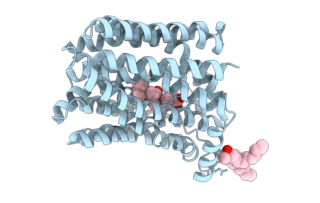
4ZP0 image
Entry Detail
PDB ID:
4ZP0
Title:
Crystal structure of E. coli multidrug transporter MdfA in complex with deoxycholate
Biological Source:
PDB Version:
Deposition Date:
2015-05-07
Release Date:
2015-08-19
Method Details:
Experimental Method:
Resolution:
2.00 Å
R-Value Free:
0.22
R-Value Work:
0.20
R-Value Observed:
0.21
Space Group:
C 1 2 1
Macromolecular Entities
Polymer Type:polypeptide(L)
Description:Multidrug transporter MdfA
Mutations:Q123R
Chain IDs:A
Chain Length:392
Number of Molecules:1
Biological Source:Escherichia coli (strain K12)
Primary Citation
Substrate-bound structure of the E. coli multidrug resistance transporter MdfA
Cell Res. 25 1060 1073 (2015)
PMID: 26238402 DOI: 10.1038/cr.2015.94

Abstact

Multidrug resistance is a serious threat to public health. Proton motive force-driven antiporters from the major facilitator superfamily (MFS) constitute a major group of multidrug-resistance transporters. Currently, no reports on crystal structures of MFS antiporters in complex with their substrates exist. The E. coli MdfA transporter is a well-studied model system for biochemical analyses of multidrug-resistance MFS antiporters. Here, we report three crystal structures of MdfA-ligand complexes at resolutions up to 2.0 Å, all in the inward-facing conformation. The substrate-binding site sits proximal to the conserved acidic residue, D34. Our mutagenesis studies support the structural observations of the substrate-binding mode and the notion that D34 responds to substrate binding by adjusting its protonation status. Taken together, our data unveil the substrate-binding mode of MFS antiporters and suggest a mechanism of transport via this group of transporters.

Legend

Protein

Chemical

Disease

Primary Citation of related structures