4ZOL image
Entry Detail
PDB ID:
4ZOL
Title:
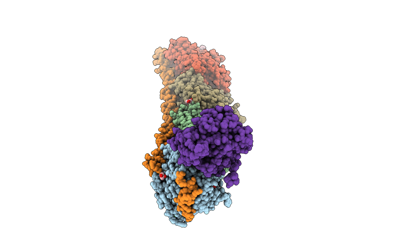
Crystal Structure of Tubulin-Stathmin-TTL-Tubulysin M Complex
Biological Source:
Host Organism:
PDB Version:
Deposition Date:
2015-05-06
Release Date:
2016-07-27
Method Details:
Experimental Method:
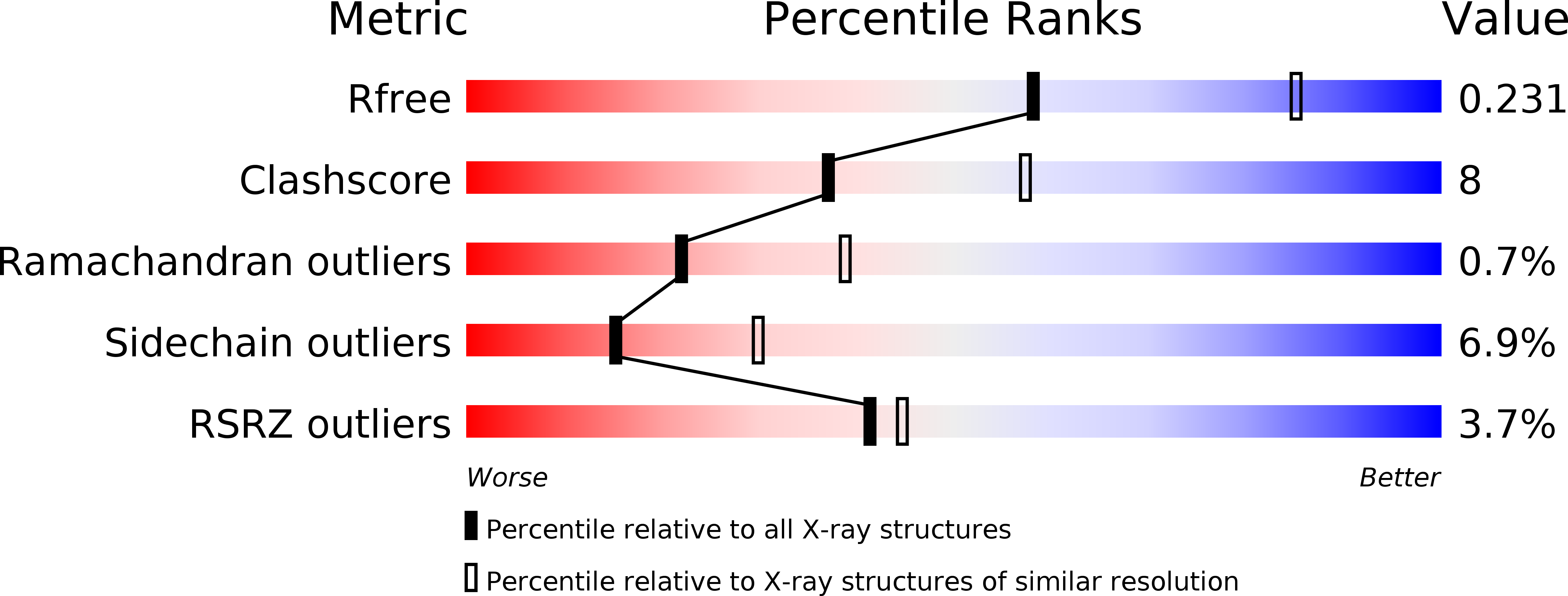
Resolution:
2.50 Å
R-Value Free:
0.23
R-Value Work:
0.17
R-Value Observed:
0.17
Space Group:
P 21 21 21
Macromolecular Entities
Polymer Type:polypeptide(L)
Description:Tubulin alpha-1B chain
Chain IDs:A, C
Chain Length:451
Number of Molecules:2
Biological Source:Sus scrofa
Polymer Type:polypeptide(L)
Description:Tubulin beta chain
Chain IDs:B, D
Chain Length:445
Number of Molecules:2
Biological Source:Sus scrofa
Polymer Type:polypeptide(L)
Description:Stathmin-4
Chain IDs:E
Chain Length:143
Number of Molecules:1
Biological Source:Rattus norvegicus
Polymer Type:polypeptide(L)
Description:Tubulin-tyrosine ligase
Chain IDs:F
Chain Length:384
Number of Molecules:1
Biological Source:Gallus gallus
Peptide-like Molecules
PRD_002237
Primary Citation
Structural Insights into the Pharmacophore of Vinca Domain Inhibitors of Microtubules
Mol.Pharmacol. 89 233 242 (2016)
PMID: 26660762 DOI: 10.1124/mol.115.100149

Abstact

Antibody-drug conjugates (ADCs) have achieved great success in cancer therapy in recent years. Some peptidyl microtubule inhibitors consisting of natural and unnatural amino acids, such as monomethyl auristatin E (MMAE) and F (MMAF), are extremely cytotoxic and have been used as a payload in ADCs. However, their precise molecular interaction with tubulin and microtubules remains unclear. We determined the crystal structures of tubulin in complex with three ultra-potent peptidyl microtubule inhibitors [MMAE, taltobulin (HTI- 286), and tubulysin M] at 2.5 Å. Our data showed that the three peptides bound to the vinca domain and shared a common and key pharmacophore containing two consecutive hydrophobic groups (Val, Ile-like side chain). These groups protruded in opposite directions into hydrophobic pockets on the tubulin β and α subunits. Nitrogen and oxygen atoms from the same backbone formed hydrogen bonds with Asn329 from the α subunit and Asp179 from the β subunit in a direction normal to the surface formed by the aforementioned hydrophobic groups. In addition, our crystal structure data indicated that tubulysin M bound to the β subunit alone, providing a structural explanation for its higher affinity. We also compared the conformations of two representative structurally different vinca domain compounds, ustiloxin D and vinblastine, with those of the aforementioned peptidyl ligands, and found that they shared a similar pharmacophore. Our findings lay a foundation for the rational design of novel vinca domain ligands and may facilitate the development of microtubule inhibitors with high specificity, affinity, and efficiency as payloads for ADCs in cancer therapy.

Legend

Protein

Chemical

Disease

Primary Citation of related structures