4ZM6 image
Deposition Date 2015-05-02
Release Date 2015-12-09
Last Version Date 2024-03-20
Entry Detail
PDB ID:
4ZM6
Title:
A unique GCN5-related glucosamine N-acetyltransferase region exist in the fungal multi-domain GH3 beta-N-acetylglucosaminidase
Biological Source:
Source Organism:
Method Details:
Experimental Method:
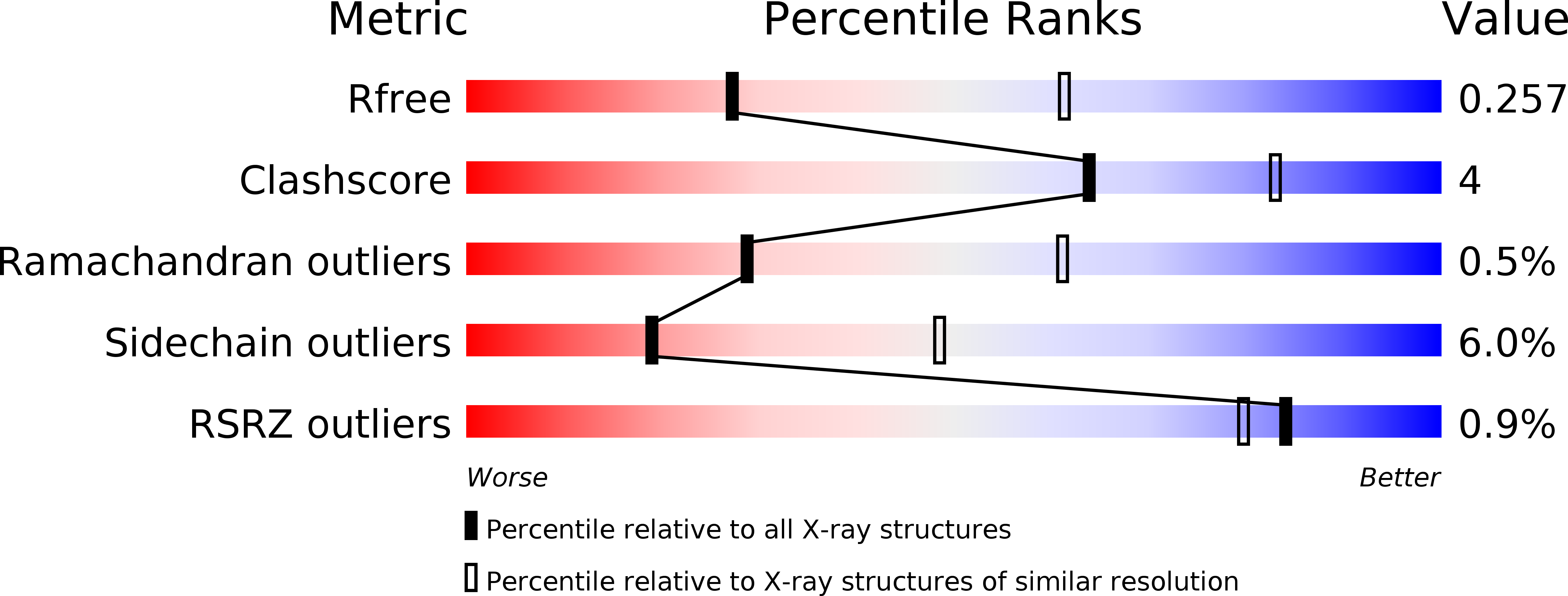
Resolution:
2.80 Å
R-Value Free:
0.25
R-Value Work:
0.22
R-Value Observed:
0.23
Space Group:
P 41 21 2
Macromolecular Entities
Structures with similar UniProt ID
Protein Blast
Polymer Type:polypeptide(L)
Molecule:N-acetyl-beta-D glucosaminidase
Gene (Uniprot):Nag
Chain IDs:A, B
Chain Length:858
Number of Molecules:2
Biological Source:Rhizomucor miehei CAU432
Primary Citation
A unique GCN5-related glucosamine N-acetyltransferase region exist in the fungal multi-domain glycoside hydrolase family 3 beta-N-acetylglucosaminidase
Sci Rep 5 18292 18292 (2015)
PMID: 26669854 DOI: 10.1038/srep18292

Abstact

Glycoside hydrolase (GH) family 3 β-N-acetylglucosaminidases widely exist in the filamentous fungi, which may play a key role in chitin metabolism of fungi. A multi-domain GH family 3 β-N-acetylglucosaminidase from Rhizomucor miehei (RmNag), exhibiting a potential N-acetyltransferase region, has been recently reported to show great potential in industrial applications. In this study, the crystal structure of RmNag was determined at 2.80 Å resolution. The three-dimensional structure of RmNag showed four distinctive domains, which belong to two distinguishable functional regions--a GH family 3 β-N-acetylglucosaminidase region (N-terminal) and a N-acetyltransferase region (C-terminal). From structural and functional analysis, the C-terminal region of RmNag was identified as a unique tandem array linking general control non-derepressible 5 (GCN5)-related N-acetyltransferase (GNAT), which displayed glucosamine N-acetyltransferase activity. Structural analysis of this glucosamine N-acetyltransferase region revealed that a unique glucosamine binding pocket is located in the pantetheine arm binding terminal region of the conserved CoA binding pocket, which is different from all known GNAT members. This is the first structural report of a glucosamine N-acetyltransferase, which provides novel structural information about substrate specificity of GNATs. The structural and functional features of this multi-domain β-N-acetylglucosaminidase could be useful in studying the catalytic mechanism of GH family 3 proteins.

Legend

Protein

Chemical

Disease

Primary Citation of related structures