
Deposition Date
2015-04-27
Release Date
2015-12-23
Last Version Date
2024-03-06
Entry Detail
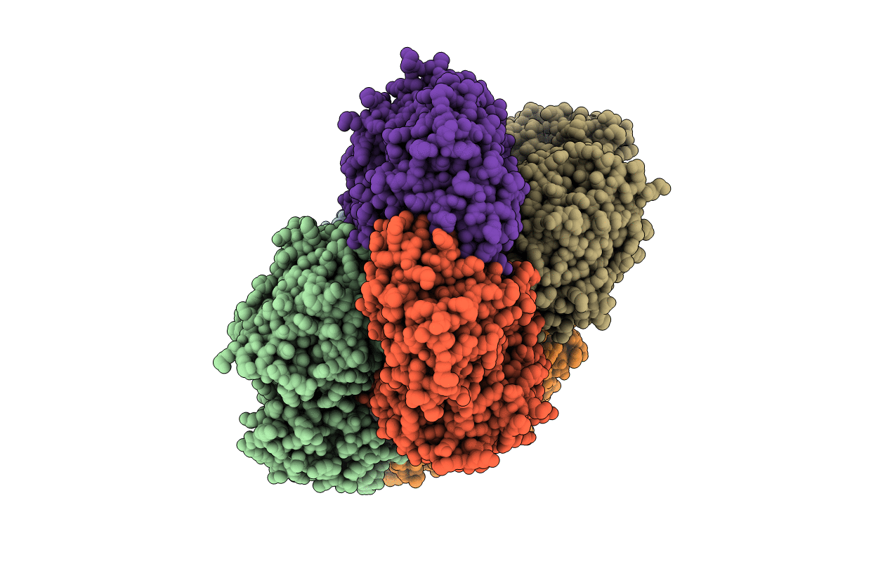
PDB ID:
4ZI6
Keywords:
Title:
Crystal structure of leucine aminopeptidase from Helicobacter pylori
Biological Source:
Source Organism(s):
Expression System(s):
Method Details:
Experimental Method:
Resolution:
2.00 Å
R-Value Free:
0.22
R-Value Work:
0.17
R-Value Observed:
0.17
Space Group:
P 1
