Deposition Date
2015-04-23
Release Date
2016-03-09
Last Version Date
2023-09-27
Entry Detail
PDB ID:
4ZGH
Keywords:
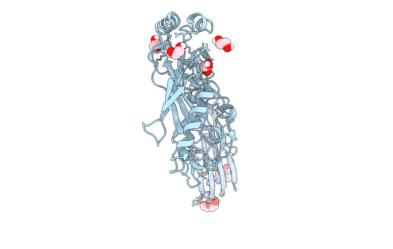
Title:
Structure of Sugar Binding Protein Pneumolysin
Biological Source:
Source Organism(s):
Streptococcus pneumoniae (Taxon ID: 373153)
Expression System(s):
Method Details:
Experimental Method:
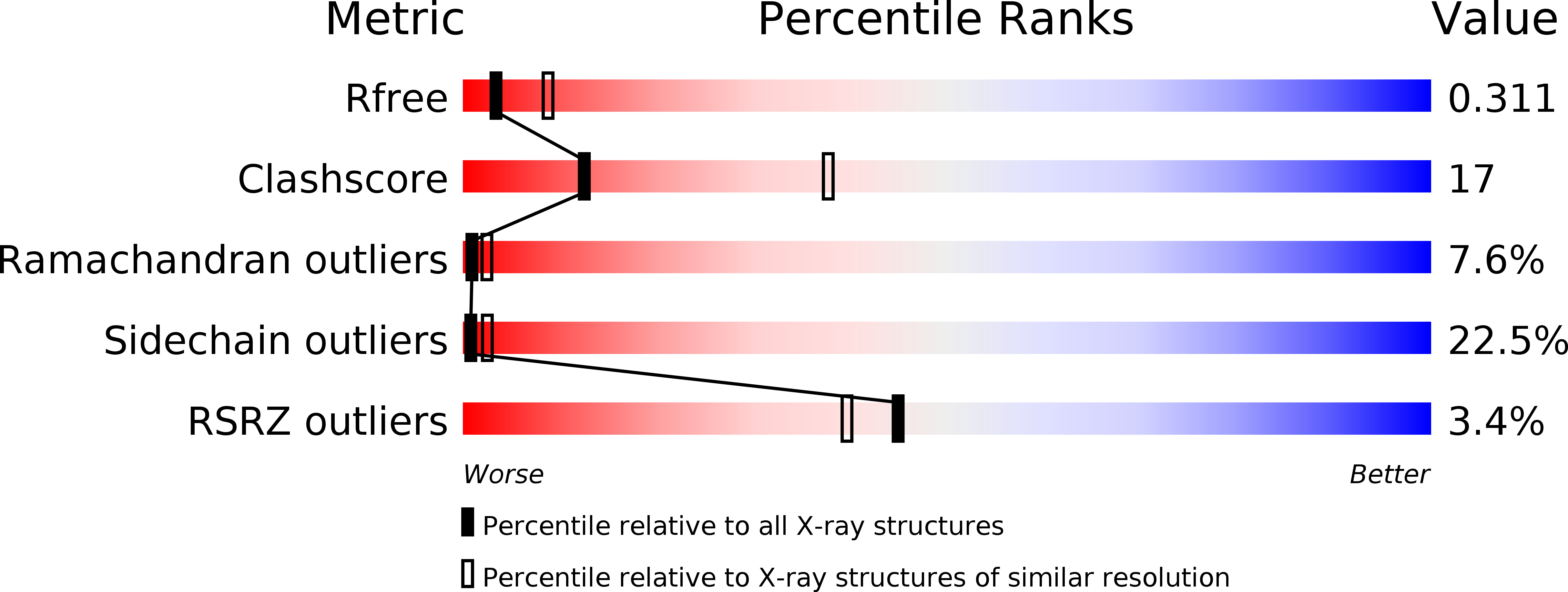
Resolution:
2.90 Å
R-Value Free:
0.30
R-Value Work:
0.19
R-Value Observed:
0.20
Space Group:
P 21 21 21