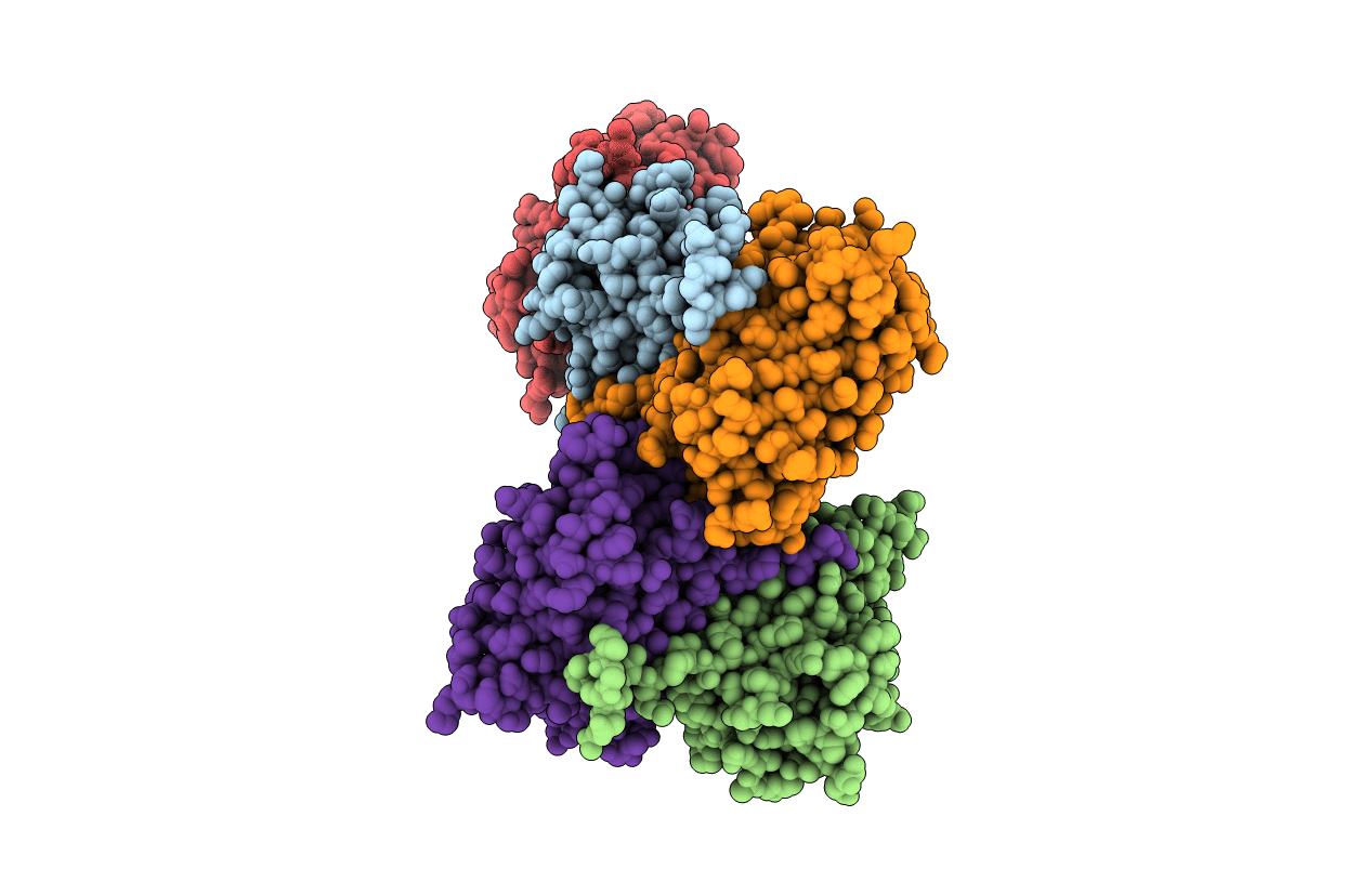
4ZFS image
Entry Detail
PDB ID:
4ZFS
Title:
Phototoxic Fluorescent Protein KillerOrange
Biological Source:
Source Organism:
Host Organism:
PDB Version:
Deposition Date:
2015-04-21
Release Date:
2015-12-23
Method Details:
Experimental Method:
Resolution:
2.01 Å
R-Value Free:
0.23
R-Value Work:
0.19
R-Value Observed:
0.19
Space Group:
C 2 2 21
Macromolecular Entities
Polymer Type:polypeptide(L)
Description:KillerOrange
Chain IDs:A, B, C, D, E
Chain Length:248
Number of Molecules:5
Biological Source:Hydrozoa
Modified Residue
Compound ID Chain ID Parent Comp ID Details 2D Image
4M9 A GLN chromophore
Primary Citation
Crystal Structure of Phototoxic Orange Fluorescent Proteins with a Tryptophan-Based Chromophore.
Plos One 10 e0145740 e0145740 (2015)
PMID: 26699366 DOI: 10.1371/journal.pone.0145740

Abstact

Phototoxic fluorescent proteins represent a sparse group of genetically encoded photosensitizers that could be used for precise light-induced inactivation of target proteins, DNA damage, and cell killing. Only two such GFP-based fluorescent proteins (FPs), KillerRed and its monomeric variant SuperNova, were described up to date. Here, we present a crystallographic study of their two orange successors, dimeric KillerOrange and monomeric mKillerOrange, at 1.81 and 1.57 Å resolution, respectively. They are the first orange-emitting protein photosensitizers with a tryptophan-based chromophore (Gln65-Trp66-Gly67). Same as their red progenitors, both orange photosensitizers have a water-filled channel connecting the chromophore to the β-barrel exterior and enabling transport of ROS. In both proteins, Trp66 of the chromophore adopts an unusual trans-cis conformation stabilized by H-bond with the nearby Gln159. This trans-cis conformation along with the water channel was shown to be a key structural feature providing bright orange emission and phototoxicity of both examined orange photosensitizers.

Legend

Protein

Chemical

Disease

Primary Citation of related structures