4ZA2 image
Deposition Date 2015-04-13
Release Date 2015-04-29
Last Version Date 2023-11-08
Entry Detail
PDB ID:
4ZA2
Keywords:
Title:
Crystal structure of Pectobacterium carotovorum 2-keto-3-deoxy-D-gluconate dehydrogenase complexed with NAD+
Biological Source:
Method Details:
Experimental Method:
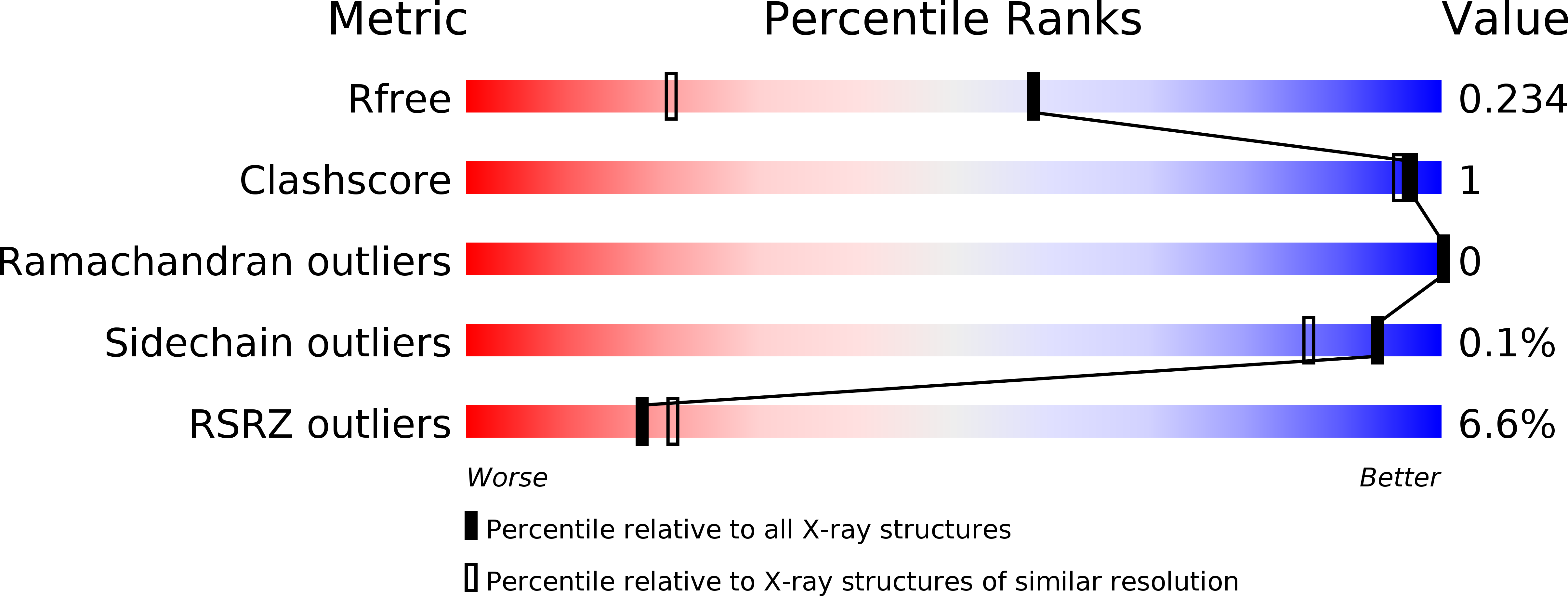
Resolution:
1.55 Å
R-Value Free:
0.22
R-Value Work:
0.19
R-Value Observed:
0.20
Space Group:
P 1 21 1
Macromolecular Entities
Polymer Type:polypeptide(L)
Molecule:2-deoxy-D-gluconate 3-dehydrogenase
Chain IDs:A, B, C, D
Chain Length:253
Number of Molecules:4
Biological Source:Pectobacterium carotovorum subsp. carotovorum
Ligand Molecules
Primary Citation
Structural determinants in bacterial 2-keto-3-deoxy-D-gluconate dehydrogenase KduD for dual-coenzyme specificity
Proteins 84 934 947 (2016)
PMID: 27028675 DOI: 10.1002/prot.25042

Abstact

Short-chain dehydrogenase/reductase (SDR) is distributed in many organisms, from bacteria to humans, and has significant roles in metabolism of carbohydrates, lipids, amino acids, and other biomolecules. An important intermediate in acidic polysaccharide metabolism is 2-keto-3-deoxy-d-gluconate (KDG). Recently, two short and long loops in Sphingomonas KDG-producing SDR enzymes (NADPH-dependent A1-R and NADH-dependent A1-R') involved in alginate metabolism were shown to be crucial for NADPH or NADH coenzyme specificity. Two SDR family enzymes-KduD from Pectobacterium carotovorum (PcaKduD) and DhuD from Streptococcus pyogenes (SpyDhuD)-prefer NADH as coenzyme, although only PcaKduD can utilize both NADPH and NADH. Both enzymes reduce 2,5-diketo-3-deoxy-d-gluconate to produce KDG. Tertiary and quaternary structures of SpyDhuD and PcaKduD and its complex with NADH were determined at high resolution (approximately 1.6 Å) by X-ray crystallography. Both PcaKduD and SpyDhuD consist of a three-layered structure, α/β/α, with a coenzyme-binding site in the Rossmann fold; similar to enzymes A1-R and A1-R', both arrange the two short and long loops close to the coenzyme-binding site. The primary structures of the two loops in PcaKduD and SpyDhuD were similar to those in A1-R' but not A1-R. Charge neutrality and moderate space at the binding site of the nucleoside ribose 2' coenzyme region were determined to be structurally crucial for dual-coenzyme specificity in PcaKduD by structural comparison of the NADH- and NADPH-specific SDR enzymes. The corresponding site in SpyDhuD was negatively charged and spatially shallow. This is the first reported study on structural determinants in SDR family KduD related to dual-coenzyme specificity. Proteins 2016; 84:934-947. © 2016 Wiley Periodicals, Inc.

Legend

Protein

Chemical

Disease

Primary Citation of related structures