4Z8N image
Entry Detail
PDB ID:
4Z8N
Keywords:
Title:
Crystal structure of the erythrocyte-binding domain from Plasmodium vivax reticulocyte-binding protein 2a (PvRBP2a)
Biological Source:
Host Organism:
PDB Version:
Deposition Date:
2015-04-09
Release Date:
2015-12-16
Method Details:
Experimental Method:
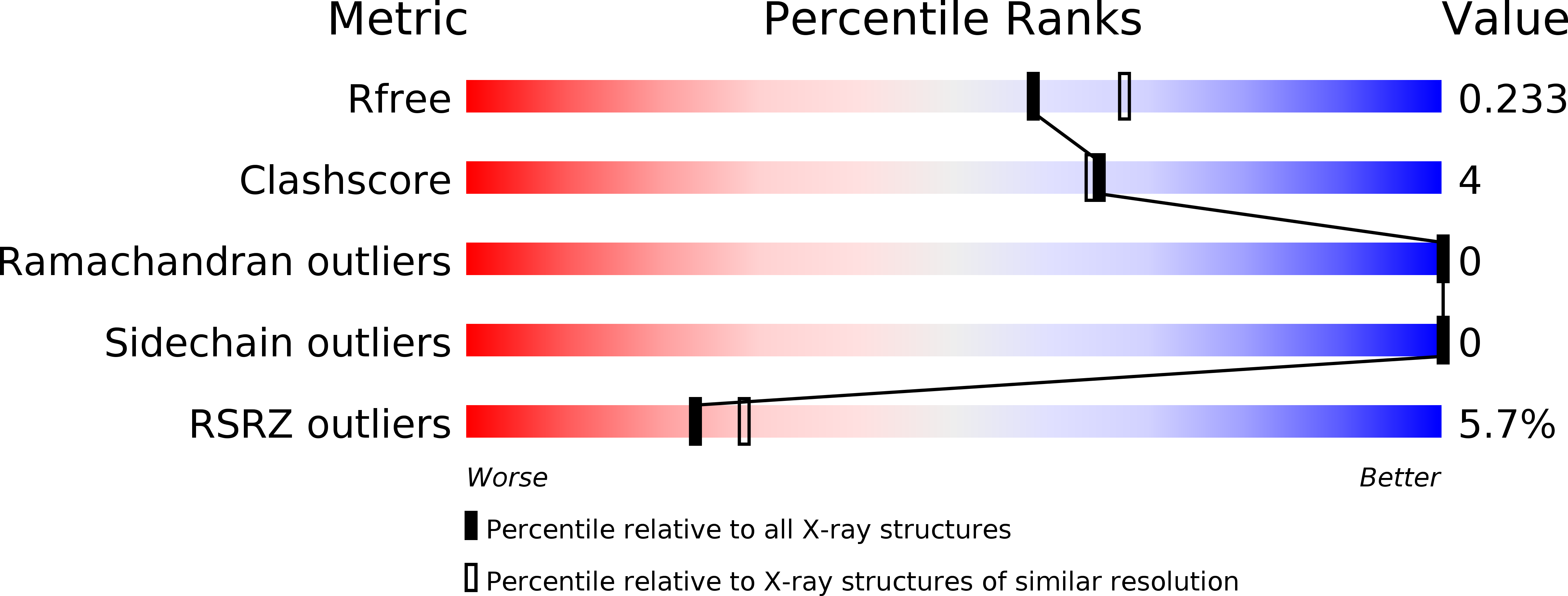
Resolution:
2.12 Å
R-Value Free:
0.22
R-Value Work:
0.20
R-Value Observed:
0.20
Space Group:
P 21 21 21
Macromolecular Entities
Polymer Type:polypeptide(L)
Description:Reticulocyte-binding protein 2a
Chain IDs:A, B
Chain Length:298
Number of Molecules:2
Biological Source:Plasmodium vivax (strain Salvador I)
Primary Citation
Structurally conserved erythrocyte-binding domain in Plasmodium provides a versatile scaffold for alternate receptor engagement.
Proc.Natl.Acad.Sci.USA 113 E191 E200 (2016)
PMID: 26715754 DOI: 10.1073/pnas.1516512113

Abstact

Understanding how malaria parasites gain entry into human red blood cells is essential for developing strategies to stop blood stage infection. Plasmodium vivax preferentially invades reticulocytes, which are immature red blood cells. The organism has two erythrocyte-binding protein families: namely, the Duffy-binding protein (PvDBP) and the reticulocyte-binding protein (PvRBP) families. Several members of the PvRBP family bind reticulocytes, specifically suggesting a role in mediating host cell selectivity of P. vivax. Here, we present, to our knowledge, the first high-resolution crystal structure of an erythrocyte-binding domain from PvRBP2a, solved at 2.12 Å resolution. The monomeric molecule consists of 10 α-helices and one short β-hairpin, and, although the structural fold is similar to that of PfRh5--the essential invasion ligand in Plasmodium falciparum--its surface properties are distinct and provide a possible mechanism for recognition of alternate receptors. Sequence alignments of the crystallized fragment of PvRBP2a with other PvRBPs highlight the conserved placement of disulfide bonds. PvRBP2a binds mature red blood cells through recognition of an erythrocyte receptor that is neuraminidase- and chymotrypsin-resistant but trypsin-sensitive. By examining the patterns of sequence diversity within field isolates, we have identified and mapped polymorphic residues to the PvRBP2a structure. Using mutagenesis, we have also defined the critical residues required for erythrocyte binding. Characterization of the structural features that govern functional erythrocyte binding for the PvRBP family provides a framework for generating new tools that block P. vivax blood stage infection.

Legend

Protein

Chemical

Disease

Primary Citation of related structures