4Z4S image
Deposition Date 2015-04-02
Release Date 2015-05-27
Last Version Date 2024-01-10
Entry Detail
PDB ID:
4Z4S
Keywords:
Title:
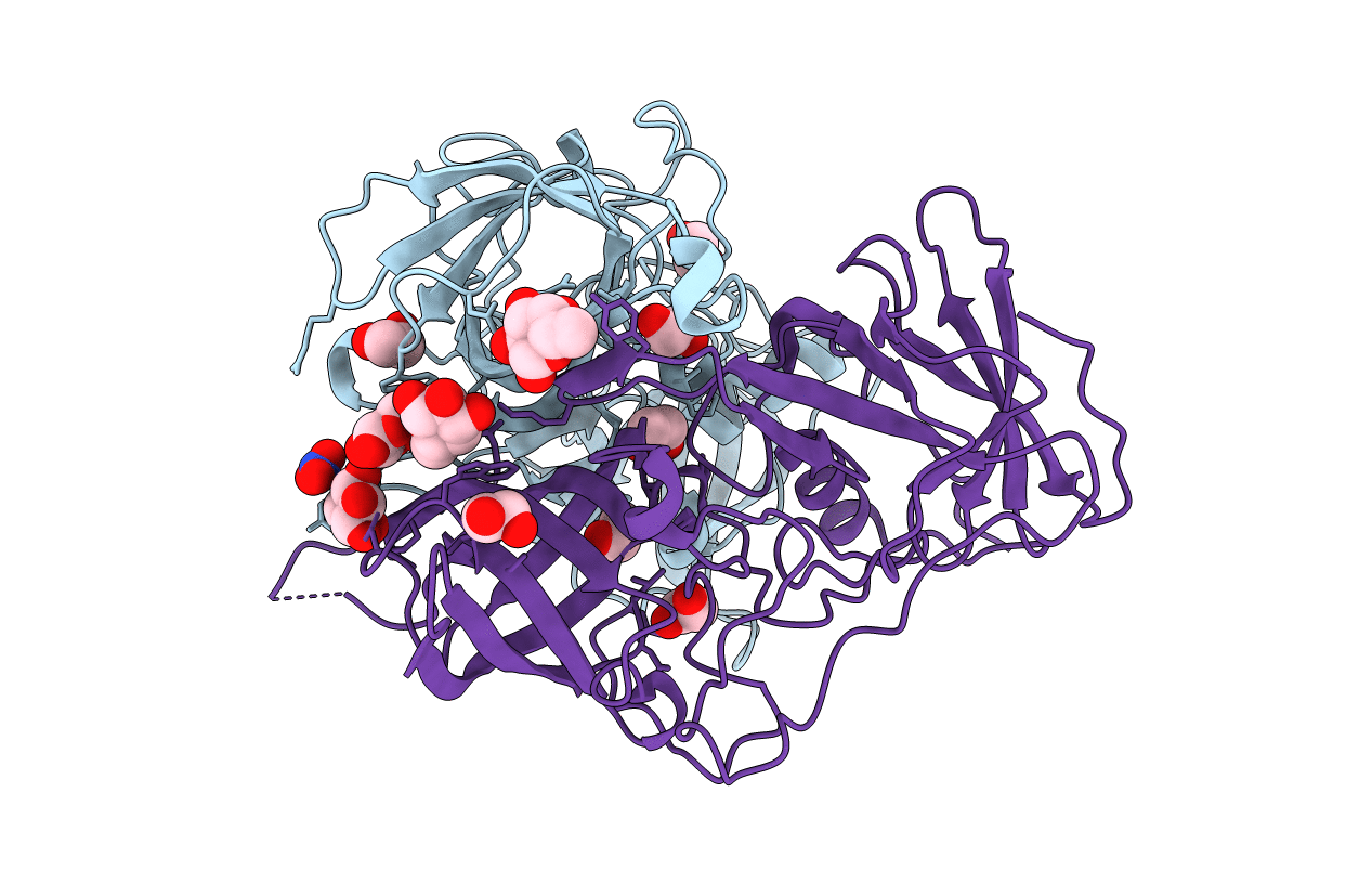
Crystal structure of GII.10 P domain in complex with 150mM fucose
Biological Source:
Source Organism:
Host Organism:
Method Details:
Experimental Method:
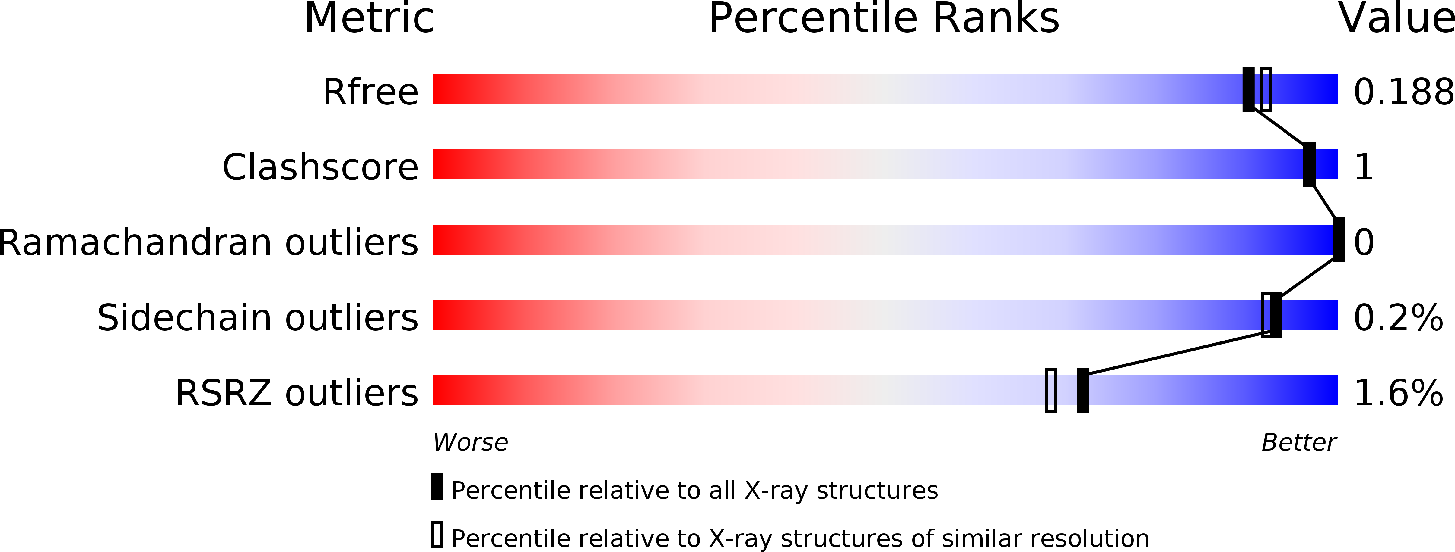
Resolution:
1.80 Å
R-Value Free:
0.18
R-Value Work:
0.14
R-Value Observed:
0.14
Space Group:
P 1 21 1
Macromolecular Entities
Polymer Type:polypeptide(L)
Molecule:Capsid protein
Chain IDs:A, B
Chain Length:315
Number of Molecules:2
Biological Source:Norovirus GII.10
Primary Citation
The sweet quartet: Binding of fucose to the norovirus capsid.
Virology 483 203 208 (2015)
PMID: 25980740 DOI: 10.1016/j.virol.2015.04.006

Abstact

Human noroviruses bind histo-blood group antigens (HBGAs) and this interaction is thought to be important for an infection. We identified two additional fucose-binding pockets (termed fucose-3/4 sites) on a genogroup II human (GII.10) norovirus-protruding (P) dimer using X-ray crystallography. Fucose-3/4 sites were located between two previously determined HBGA binding pockets (termed fucose-1/2 sites). We found that four fucose molecules were capable of binding altogether at fucose-1/2/3/4 sites on the P dimer, though the fucose molecules bound in a dose-dependent and step-wise manner. We also showed that HBGA B-trisaccharide molecules bound in a similar way at the fucose-1/2 sites. Interestingly, we discovered that the monomers of the P dimer were asymmetrical in an unliganded state and when a single B-trisaccharide molecule bound, but were symmetrical when two B-trisaccharide molecules bound. We postulate that the symmetrical dimers might favor HBGA binding interactions at fucose-1/2 sites.

Legend

Protein

Chemical

Disease

Primary Citation of related structures