4Z3E image
Entry Detail
PDB ID:
4Z3E
Title:
Crystal structure of the lectin domain of PapG from E. coli BI47 in complex with SSEA4 in space group P212121
Biological Source:
Source Organism:
PDB Version:
Deposition Date:
2015-03-31
Release Date:
2016-04-13
Method Details:
Experimental Method:
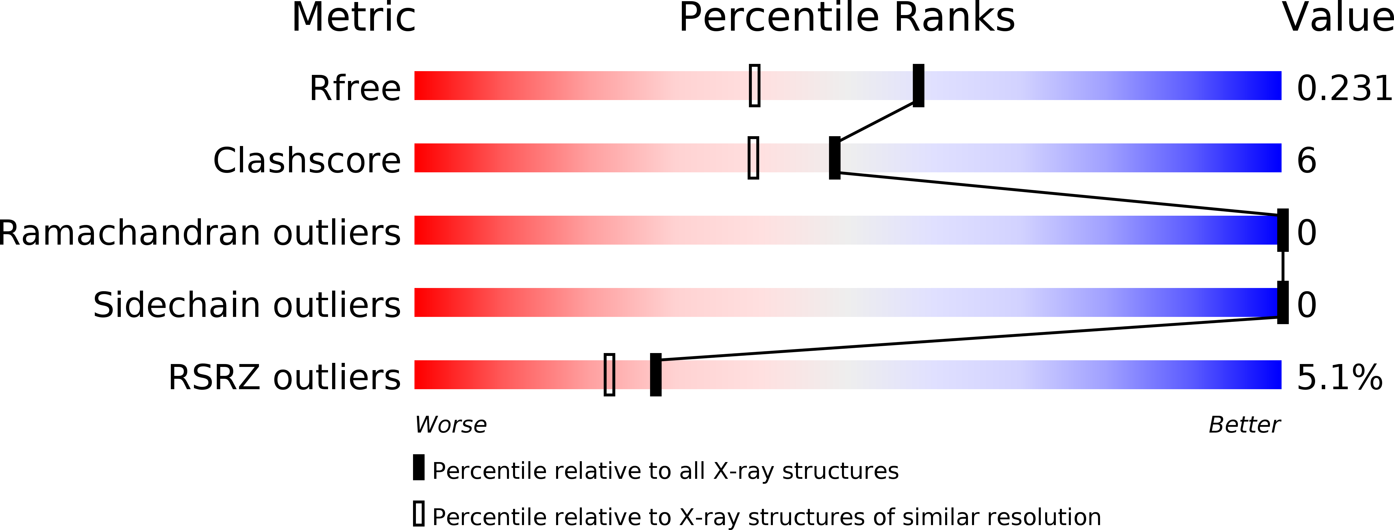
Resolution:
1.80 Å
R-Value Free:
0.22
R-Value Work:
0.17
R-Value Observed:
0.18
Space Group:
P 21 21 21
Macromolecular Entities
Polymer Type:polypeptide(L)
Description:PapG, lectin domain
Chain IDs:A
Chain Length:198
Number of Molecules:1
Biological Source:ESCHERICHIA COLI
Primary Citation
Carbohydrate-Lectin Interactions: An Unexpected Contribution to Affinity.
Chembiochem 18 539 544 (2017)
PMID: 28076665 DOI: 10.1002/cbic.201600615

Abstact

Uropathogenic E. coli exploit PapG-II adhesin for infecting host cells of the kidney; the expression of PapG-II at the tip of bacterial pili correlates with the onset of pyelonephritis in humans, a potentially life-threatening condition. It was envisaged that blocking PapG-II (and thus bacterial adhesion) would provide a viable therapeutic alternative to conventional antibiotic treatment. In our search for potent PapG-II antagonists, we observed an increase in affinity when tetrasaccharide 1, the natural ligand of PapG-II in human kidneys, was elongated to hexasaccharide 2, even though the additional Siaα(2-3)Gal extension is not in direct contact with the lectin. ITC studies suggest that the increased affinity results from partial desolvation of nonbinding regions of the hexasaccharide; this is ultimately responsible for perturbation of the outer hydration layers. Our results are in agreement with previous observations and suggest a general mechanism for modulating carbohydrate-protein interactions based on nonbinding regions of the ligand.

Legend

Protein

Chemical

Disease

Primary Citation of related structures