4Z07 image
Deposition Date 2015-03-25
Release Date 2016-04-13
Last Version Date 2023-09-27
Entry Detail
PDB ID:
4Z07
Keywords:
Title:
Co-crystal structure of the tandem CNB (CNB-A/B) domains of human PKG I beta with cGMP
Biological Source:
Source Organism:
Homo sapiens (Taxon ID: 9606)
Host Organism:
Method Details:
Experimental Method:
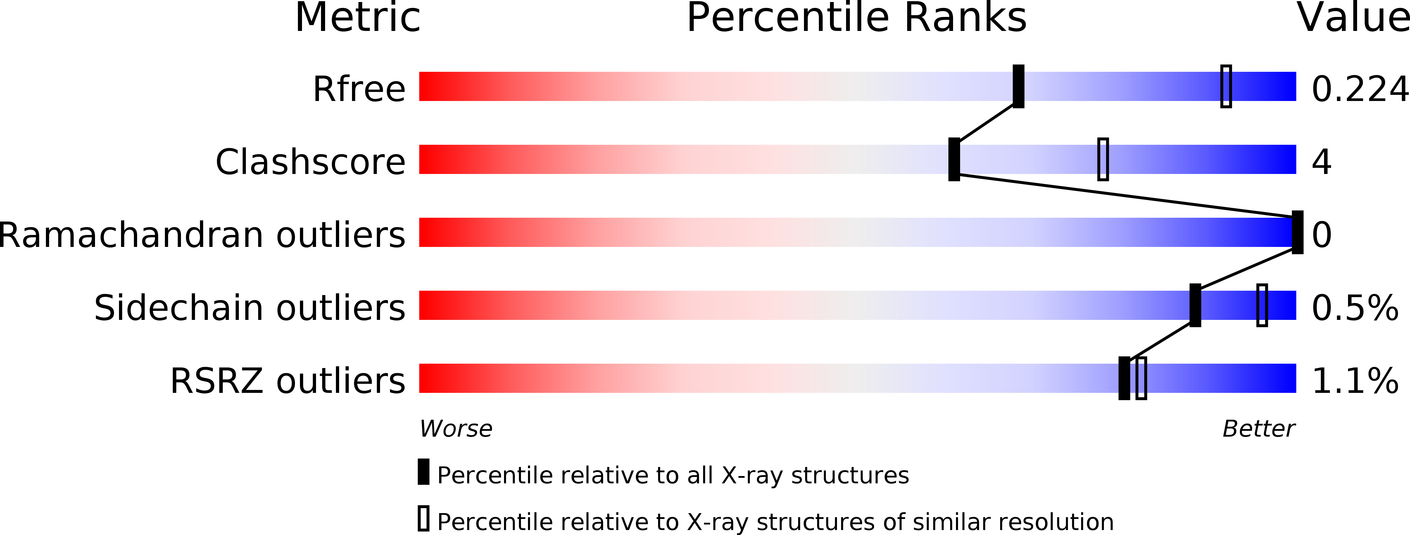
Resolution:
2.50 Å
R-Value Free:
0.22
R-Value Work:
0.16
R-Value Observed:
0.16
Space Group:
C 2 2 2
Macromolecular Entities
Structures with similar UniProt ID
Protein Blast
Polymer Type:polypeptide(L)
Molecule:cGMP-dependent protein kinase 1
Gene (Uniprot):PRKG1
Chain IDs:A, B (auth: C), C (auth: E)
Chain Length:262
Number of Molecules:3
Biological Source:Homo sapiens
Primary Citation
Crystal Structure of PKG I:cGMP Complex Reveals a cGMP-Mediated Dimeric Interface that Facilitates cGMP-Induced Activation.
Structure 24 710 720 (2016)
PMID: 27066748 DOI: 10.1016/j.str.2016.03.009

Abstact

Cyclic guanosine monophosphate (cGMP)-dependent protein kinase (PKG) is a key regulator of smooth muscle and vascular tone and represents an important drug target for treating hypertensive diseases and erectile dysfunction. Despite its importance, its activation mechanism is not fully understood. To understand the activation mechanism, we determined a 2.5 Å crystal structure of the PKG I regulatory (R) domain bound with cGMP, which represents the activated state. Although we used a monomeric domain for crystallization, the structure reveals that two R domains form a symmetric dimer where the cGMP bound at high-affinity pockets provide critical dimeric contacts. Small-angle X-ray scattering and mutagenesis support this dimer model, suggesting that the dimer interface modulates kinase activation. Finally, structural comparison with the homologous cyclic AMP-dependent protein kinase reveals that PKG is drastically different from protein kinase A in its active conformation, suggesting a novel activation mechanism for PKG.

Legend

Protein

Chemical

Disease

Primary Citation of related structures