Deposition Date
2015-03-25
Release Date
2015-05-13
Last Version Date
2024-11-20
Entry Detail
PDB ID:
4YZS
Keywords:
Title:
Crystal structures reveal transient PERK luminal domain tetramerization in ER stress signaling
Biological Source:
Source Organism(s):
Homo sapiens (Taxon ID: 9606)
Expression System(s):
Method Details:
Experimental Method:
Resolution:
3.14 Å
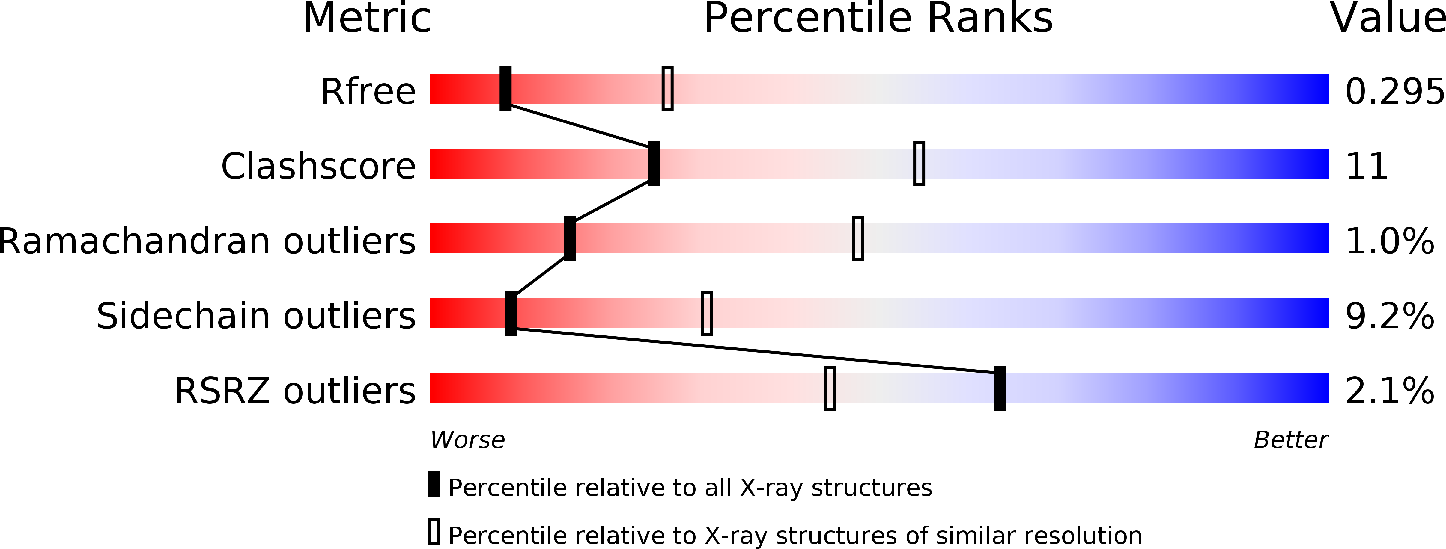
R-Value Free:
0.28
R-Value Work:
0.23
R-Value Observed:
0.24
Space Group:
P 41 21 2