
Deposition Date
2015-03-22
Release Date
2015-09-23
Last Version Date
2024-11-06
Entry Detail
PDB ID:
4YX2
Keywords:
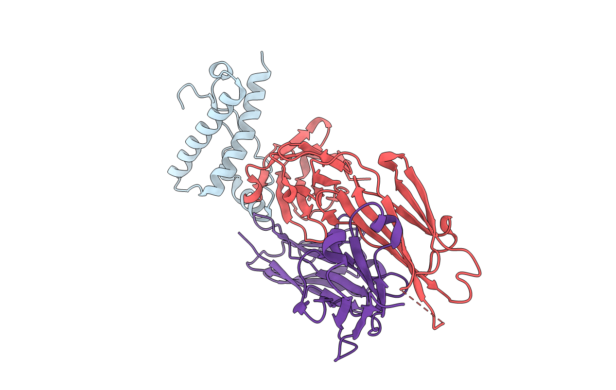
Title:
Crystal structure of Bovine prion protein complexed with POM1 FAB
Biological Source:
Source Organism(s):
Bos taurus (Taxon ID: 9913)
Mus musculus (Taxon ID: 10090)
Mus musculus (Taxon ID: 10090)
Expression System(s):
Method Details:
Experimental Method:
Resolution:
2.19 Å
R-Value Free:
0.26
R-Value Work:
0.21
R-Value Observed:
0.21
Space Group:
C 1 2 1
